Most people have heard of arthritis, lupus, or osteoporosis, but relatively few recognize the name Ehlers-Danlos syndrome (EDS). Yet this group of inherited connective tissue disorders affects thousands of people worldwide and can profoundly alter nearly every aspect of daily life. For some, it means unusually flexible joints and chronic pain. For others, it carries the risk of ruptured blood vessels or internal organs. Between these extremes lies a remarkably diverse family of disorders that illustrates just how important the body’s structural framework truly is.
Ehlers-Danlos syndrome is not a single disease but a collection of thirteen recognized hereditary disorders that share one common feature: abnormalities in connective tissue. Connective tissue forms the body’s internal scaffolding, providing strength, flexibility, and support to the skin, joints, blood vessels, ligaments, tendons, and many internal organs. When this framework is weakened, the effects can extend throughout the body.
Although EDS has long been considered rare, growing awareness and improved diagnostic methods suggest it may be more common than previously believed. Many individuals spend years seeking an explanation for symptoms that are dismissed as unrelated or attributed to anxiety, chronic pain syndromes, or simple clumsiness. Increased recognition has begun shortening this diagnostic journey, but many patients still experience significant delays before receiving an accurate diagnosis.
The Biology Behind EDS
At the heart of Ehlers-Danlos syndrome is collagen, the most abundant protein in the human body. Collagen functions much like the steel reinforcing bars embedded within concrete. It gives tissues strength while allowing them to remain flexible enough to move without tearing.
Collagen is found almost everywhere, including the skin, tendons, ligaments, cartilage, bones, blood vessels, intestines, and the protective coverings surrounding many organs. Multiple genes direct the production, assembly, and maintenance of collagen fibers and other components of the extracellular matrix—the microscopic framework that supports cells throughout the body.
In EDS, inherited genetic mutations interfere with this process. Depending on which gene is affected, collagen may be produced incorrectly, assembled improperly, or fail to provide adequate structural support. The resulting weakness affects whichever tissues rely most heavily on the defective protein.
The pattern of inheritance varies among the different forms of EDS. Many are inherited in an autosomal dominant fashion, meaning that a child has a 50 percent chance of inheriting the disorder if one parent carries the abnormal gene. Other forms are autosomal recessive, requiring defective copies from both parents, while some cases result from entirely new mutations with no previous family history.
One notable exception remains the most common subtype: hypermobile Ehlers-Danlos syndrome (hEDS). Despite years of intensive research, no single genetic mutation has yet been identified. As a result, hEDS remains a clinical diagnosis based on characteristic symptoms and examination findings rather than laboratory confirmation. Identifying its underlying genetic basis is one of the major goals of current EDS research.
The syndrome bears the names of two physicians who helped characterize it in the early twentieth century: Danish dermatologist Edvard Ehlers and French physician Henri-Alexandre Danlos.
A Family of Disorders
The 2017 International Classification recognizes thirteen distinct forms of Ehlers-Danlos syndrome. While all involve connective tissue abnormalities, each subtype has its own characteristic pattern of symptoms and complications.
Hypermobile EDS
Hypermobile EDS is by far the most frequently diagnosed subtype. Its defining features include generalized joint hypermobility, chronic musculoskeletal pain, and varying degrees of skin involvement. Individuals may be able to bend their fingers, elbows, knees, or spine far beyond the normal range of motion.
Although hypermobility can appear impressive, it often comes at a significant cost. Joints that move too freely lack stability, making them prone to sprains, partial dislocations, and complete dislocations during everyday activities. Chronic pain and fatigue frequently become the most disabling aspects of the disorder.
Many people with hypermobile EDS also experience additional conditions that appear to occur more frequently than expected. These include disorders of the autonomic nervous system, particularly postural orthostatic tachycardia syndrome (POTS), gastrointestinal motility problems, chronic headaches, and persistent fatigue. Researchers continue to investigate why these conditions commonly occur together.
Classical EDS
Classical EDS primarily affects the skin and joints. It usually results from mutations involving genes responsible for type V collagen.
Patients typically have remarkably soft, velvety skin that stretches much farther than normal before returning to its original position. Wounds often heal poorly, producing thin, widened scars sometimes described as “cigarette-paper scars.” Easy bruising is common, and even relatively minor injuries may leave permanent marks.
Joint hypermobility is also a prominent feature, although the degree varies considerably from person to person.
Vascular EDS
Among all forms of Ehlers-Danlos syndrome, vascular EDS is the most serious. It results from mutations affecting type III collagen, an important structural protein within blood vessel walls and many internal organs.
Unlike the dramatic flexibility often seen in hypermobile EDS, patients with vascular EDS may have only mild joint laxity, usually involving the small joints of the hands and feet. Instead, the major concern is tissue fragility.
Arteries may develop aneurysms, tears, or spontaneous ruptures. The intestines and uterus are also unusually fragile, creating life-threatening medical emergencies that often occur without warning.
Many individuals with vascular EDS also have recognizable physical characteristics, including thin translucent skin, prominent veins, a narrow nose, thin lips, and relatively large, prominent eyes. These findings, however, are not present in every patient.
The Rarer Forms
The remaining subtypes are uncommon, with some documented in only a small number of families worldwide.
Kyphoscoliotic EDS often presents during infancy with severe muscle weakness and progressive curvature of the spine. Arthrochalasia EDS causes profound joint instability beginning at birth, while dermatosparaxis EDS produces exceptionally fragile, sagging skin that tears easily.
Other rare forms primarily affect the eyes, heart valves, muscles, teeth, or periodontal tissues. Although individually uncommon, these disorders illustrate how connective tissue supports virtually every organ system in the body.
One Disease, Many Presentations
One of the greatest challenges in understanding Ehlers-Danlos syndrome is its extraordinary variability. Even individuals carrying the same genetic mutation may experience dramatically different symptoms and levels of disability. Some remain physically active with relatively mild limitations, while others develop chronic pain, repeated joint injuries, or life-threatening vascular complications.
This wide spectrum often complicates diagnosis. Physicians unfamiliar with EDS may not immediately recognize that seemingly unrelated symptoms—frequent sprains, digestive problems, easy bruising, chronic fatigue, headaches, and dizziness—can all stem from a single underlying connective tissue disorder.
Ehlers-Danlos syndrome occupies a peculiar position in medicine: common enough that most physicians will encounter it, but rare enough — and complex enough — that many will misidentify it or underestimate it. As awareness grows and genetic testing becomes more accessible, diagnostic delays are slowly shortening.
For patients living with EDS today, the message is mixed but not without hope. For most, a normal lifespan is likely, though the journey will require proactive self-advocacy, knowledgeable medical partnerships, and real-world adaptation. For those with vascular EDS, the risks are serious and demand vigilance — but even here, the medical community is making progress.
Recognizing these patterns is the first step toward appropriate treatment. Although EDS cannot currently be cured, an accurate diagnosis allows patients and physicians to anticipate complications, tailor therapy, and improve long-term quality of life.
The information provided in this article is intended for general educational and informational purposes only and does not constitute medical advice. It should not be used as a substitute for professional medical advice, diagnosis, or treatment.
Always seek the guidance of a qualified healthcare provider with any questions you may have regarding a medical condition or treatment. Never disregard professional medical advice or delay seeking it because of something you have read here.
If you are experiencing a medical emergency, call 911 or your local emergency number immediately.
The author of this article is a licensed physician, but the views expressed here are solely those of the author and do not represent the official position of any hospital, health system, or medical organization with which the author may be affiliated.
Illustration Generated by author using ChatGPT.
Sources and Further Reading:
Note: The 2017 diagnostic classification is the current standard; updated criteria are expected in 2026.
Structure, Function, Testing, and Clinical Implications
If you’ve ever had blood drawn during a bout of illness or a workup for heart disease risk, there’s a decent chance your doctor ordered a C-reactive protein test, usually shortened to CRP. It’s one of the most widely ordered lab tests in the world. CRP is one of the body’s most sensitive early-warning systems for inflammation. Understanding where it comes from, what it does, and how doctors use it reveals a lot about how the immune system operates and why chronic low-grade inflammation has become such a central concern in modern medicine. Warning: doctor talk will follow.
A Protein With a Peculiar Origin Story
CRP was discovered almost by accident in 1930 by William Tillett and Thomas Francis, working in Oswald Avery’s laboratory at Rockefeller University. They were studying patients with acute pneumococcal pneumonia caused by the bacteria Streptococcus pneumoniae and noticed that blood from acutely ill patients caused a specific bacterial substance to clump together. That substance was the “C polysaccharide” in the bacterial cell wall, which gave the protein its name.
For decades CRP was used as a crude yes-or-no indicator of serious infection. It wasn’t until the late 20th century that more sensitive lab techniques revealed its value for detecting the subtle, chronic inflammation now linked to cardiovascular disease and opening a major new chapter in preventive medicine. I had been in practice for several years before I even became aware of CRP.
Shape Matters: The Five-Sided Structure of CRP
CRP belongs to a protein family called the pentraxins, named from the Greek word for “five” because of the protein’s pentagonal shape. Under an electron microscope, CRP looks like five identical subunits arranged in a ring, roughly like five coins fanned into a disc. For history buffs, it resembles the five-star rank worn by General Eisenhower.
Each subunit carries a binding site made of two calcium ions nestled next to a small hydrophobic pocket. This is where CRP does its recognition work: the calcium-dependent sites allow it to grab onto phosphocholine (PC), a molecule found in the membranes of damaged and dying cells, as well as in the outer coatings of many bacteria and fungi. Phosphocholine is so widespread in biology that this single binding chemistry lets CRP respond to an enormous range of threats including microbial invaders and the body’s own dead cells.
CRP doesn’t always exist in this five-subunit ring form. The pentameric version (pCRP) circulates in healthy blood. But at sites of active tissue damage, such as inside an inflamed artery wall, it can break apart into individual monomers (mCRP). These two forms turn out to have strikingly different effects on the immune system.
What CRP Does
It’s tempting to think of CRP as a passive number on a lab report, but it’s actually an active participant in the immune response. It plays at least four major roles.
First, CRP is a pattern recognition molecule. Unlike the antibodies generated by your adaptive immune system which take days or weeks to respond to a specific threat, CRP responds immediately to general molecular patterns shared by pathogens and damaged cells. This makes it a first-responder tool within the innate immune system, the body’s rapid defense network.
Second, once CRP binds to a pathogen or dying cell, it acts as an opsonin, essentially a molecular flag that says “eat this.” Immune cells called phagocytes (macrophages and neutrophils) recognize CRP-coated targets and engulf them, accelerating the clearance of both bacteria and cellular debris.
Third, CRP can activate the complement system, a cascade of proteins that further tags pathogens for destruction. Crucially, CRP appears to stop short of triggering the most destructive steps of that cascade. The net effect is a measured, targeted immune response rather than an all-out inflammatory assault.
Fourth, and this is where things get interesting, the pentameric and monomeric forms of CRP have opposing effects. The pentameric form in circulation is primarily anti-inflammatory, quietly clearing dead cells without triggering unnecessary immune activation. But the monomeric form that appears at sites of tissue damage can amplify inflammation: activating platelets, recruiting immune cells to vessel walls, and stimulating the release of the inflammatory signaling molecule. CRP is neither simply good nor simply bad — it’s context-dependent, functioning as a careful janitor in healthy tissue but potentially fanning the flames in already-inflamed environments.
Fast, Sensitive, and Liver-Made
CRP is produced primarily by liver cells (hepatocytes) in response to immune signals. When immune cells detect infection or tissue damage, they release cytokines, which travel to the liver and switch on CRP production.
One of CRP’s great clinical virtues is speed. Levels can begin rising within four to six hours of an inflammatory event and typically peak within 24 to 48 hours. When the inflammation resolves, CRP falls just as quickly, its half-life in circulation is only about 19 hours. This fast-on, fast-off behavior makes CRP an excellent real-time readout of what the immune system is doing right now, not weeks ago. Because of this early availability, it is referred to as an acute phase reactant.
Under normal, non-inflammatory conditions, CRP in the blood typically measures below 1 milligram per liter (mg/L). During serious bacterial infection or major tissue injury, levels can spike above 400 mg/L — a several-hundred-fold increase. CRP also has a practical advantage for testing: it doesn’t fluctuate throughout the day, and fasting is not required before a blood draw.
The Many Uses of CRP in Clinical Medicine
The oldest application of CRP testing is detecting and monitoring bacterial infection. Very high values — generally above 100 mg/L — strongly suggest bacterial rather than viral infection, since viruses tend to provoke a much more modest CRP rise. In sepsis (the life-threatening systemic response to infection), CRP is one of several markers used to assess severity and track the response to treatment.
CRP is also routinely used to monitor autoimmune diseases. In rheumatoid arthritis, CRP is included in standard disease activity scoring tools. Tracking CRP over time helps clinicians judge whether treatments like disease-modifying drugs are working. One notable exception: in systemic lupus erythematosus (SLE), CRP often stays surprisingly low even during active flares.
Perhaps the most debated expansion of CRP testing in recent decades is its role in cardiovascular risk assessment. That story begins with the recognition that atherosclerosis, the plaque buildup inside artery walls, is fundamentally an inflammatory process, not just a plumbing problem caused by too much cholesterol. As that insight took hold in the 1990s, researchers began asking whether CRP could predict heart attack risk the way cholesterol does.
The answer was yes, but only with a more sensitive test. Standard CRP assays can’t detect levels below about 10 mg/L, which is fine for infections but misses the chronic low-level inflammation relevant to cardiovascular risk. A newer high-sensitivity CRP test (hs-CRP) can measure levels as low as 0.01 mg/L. Using hs-CRP, researchers found that even modest CRP elevations, within the range once considered entirely normal, carry meaningful cardiovascular risk.
The American Heart Association and the CDC use these hs-CRP thresholds for cardiovascular risk: below 1 mg/L is low risk; 1 to 3 mg/L is intermediate risk; above 3 mg/L is higher risk. A large UK Biobank analysis of nearly 450,000 people found that individuals with hs-CRP above 3 mg/L had a 34% higher risk of major cardiovascular events and a 61% higher risk of cardiovascular death compared to those below 1 mg/L.
The landmark JUPITER trial demonstrated that patients with low LDL cholesterol, but elevated hs-CRP (above 2 mg/L) still benefited substantially from statin therapy — in terms of reduced heart attacks and strokes. This reframed how cardiologists think about inflammation as a cardiovascular target independent of cholesterol levels.
Beyond infection, autoimmune disorders, and heart disease, elevated CRP has been linked to type 2 diabetes, metabolic syndrome, chronic kidney disease, COPD, depression, and neurodegenerative diseases, all conditions where chronic low-grade inflammation is increasingly recognized as a contributing factor rather than just a side effect.
Measuring CRP: The Standard Test vs. the High-Sensitivity Test
CRP testing is straightforward, just a standard blood draw, no fasting required. Modern automated lab analyzers make it fast and inexpensive, which explains why it shows up so often in clinical workups.
The standard CRP test is the workhorse for detecting acute infection and monitoring inflammatory flares in hospitalized patients. The hs-CRP test uses more sensitive techniques and is the test used for cardiovascular risk assessment.
Because hs-CRP can fluctuate modestly from day to day, cardiovascular guidelines recommend averaging two measurements taken about two weeks apart. Acute illness, recent injury, or even a hard workout can temporarily elevate hs-CRP and produce a misleading result.
Several factors can push CRP higher independently of the disease in question: obesity, smoking, high blood pressure, metabolic syndrome, low HDL cholesterol, and chronic low-grade infections such as gum disease. Age also gradually raises baseline CRP. On the other side, moderate physical activity, weight loss, and statins are all associated with lower CRP — which partly explains the broader cardiovascular benefits of statin therapy beyond cholesterol reduction.
What CRP Can’t Tell You
For all its usefulness, CRP is a nonspecific marker. A reading of 50 mg/L is consistent with a kidney infection, an autoimmune flare, a recent heart attack, or an abdominal cancer. CRP tells you that inflammation or tissue damage is happening somewhere; it doesn’t tell you where or why. Clinical context including symptoms, medical history, and other lab results, is essential for interpretation.
There’s also a genuine unresolved debate about whether CRP elevation actually causes cardiovascular disease or is simply a marker of underlying inflammatory risk. Animal studies in CRP-deficient or CRP-enhanced models have produced inconsistent results. Mendelian randomization studies in humans, a statistical technique that uses genetic variants to approximate a randomized experiment, have generally not supported CRP as a causal driver of heart disease, suggesting it may be more of a reaction than a root cause.
Finally, hs-CRP testing, despite strong evidence, remains underused in primary care, particularly in primary prevention. The 2024 European Society of Cardiology guidelines for chronic coronary syndromes did recommend assessing hs-CRP in patients with suspected coronary artery disease, which reflects accumulating evidence for its utility, but broader implementation lags behind the science. I have to admit, when I was still in active practice, I was unaware of the role of hs-CRP in primary prevention. At the time, it was generally thought to be of use in secondary prevention—actions taken after an initial event. Be sure and ask your doctor about it.
CRP is, in the end, a remarkably versatile tool — a protein that has been doing immune surveillance since long before medicine had a name for it, and one that continues to find new clinical relevance with each decade of research.
Medical Disclaimer
The information provided in this article is intended for general educational and informational purposes only and does not constitute medical advice. It should not be used as a substitute for professional medical advice, diagnosis, or treatment.
Always seek the guidance of a qualified healthcare provider with any questions you may have regarding a medical condition or treatment. Never disregard professional medical advice or delay seeking it because of something you have read here.
If you are experiencing a medical emergency, call 911 or your local emergency number immediately.
The author of this article is a licensed physician, but the views expressed here are solely those of the author and do not represent the official position of any hospital, health system, or medical organization with which the author may be affiliated.
Image generated by the author using ChatGPT
Sources
The following peer-reviewed and clinical references support this article:
Zhou et al. (2024). C-reactive protein: structure, function, regulation, and role in clinical diseases. Frontiers in Immunology. — https://pmc.ncbi.nlm.nih.gov/articles/PMC11211361/
Agrawal & Wu (2024). Editorial: Biology of C-reactive protein. Frontiers in Immunology. — https://www.frontiersin.org/journals/immunology/articles/10.3389/fimmu.2024.1445001/full
Volanakis (2001). Human C-reactive protein: expression, structure, and function. Molecular Immunology. PubMed. — https://pubmed.ncbi.nlm.nih.gov/11532280/
Thompson et al. (2004). C-reactive protein. Journal of Biological Chemistry. — https://www.jbc.org/article/S0021-9258(19)32228-8/fulltext
Salazar et al. (2014). C-Reactive Protein: An In-Depth Look into Structure, Function, and Regulation. International Scholarly Research Notices. PMC. — https://www.ncbi.nlm.nih.gov/pmc/articles/PMC4897210/
StatPearls: C-Reactive Protein: Clinical Relevance and Interpretation. PubMed. — https://pubmed.ncbi.nlm.nih.gov/28722873/
Labcorp: C-Reactive Protein (CRP), High Sensitivity (Cardiac Risk Assessment). — https://www.labcorp.com/tests/120766/c-reactive-protein-crp-high-sensitivity-cardiac-risk-assessment
Larsen et al. (2024). C-reactive protein and cardiovascular risk in the general population. European Heart Journal. — https://academic.oup.com/eurheartj/advance-article/doi/10.1093/eurheartj/ehaf937/8377304
Shetty & Kawaguchi (2024). Role of CRP in disease progression, diagnosis and management. PMC. — https://pmc.ncbi.nlm.nih.gov/articles/PMC11569793/
McDonnell et al. (2024). C-reactive protein complement-ary structures in RA. Elsevier/Immunology Letters. — https://www.binasss.sa.cr/ago24/21.pdf
CRP kinetics as predictor in immune checkpoint inhibitor therapy. BJC Reports / Nature. — https://www.nature.com/articles/s44276-023-00005-x
Borges et al. (2024). Battle of the Biomarkers of Systemic Inflammation (CRP vs cfDNA). PMC. — https://pmc.ncbi.nlm.nih.gov/articles/PMC12024891/
Assay Genie: C-reactive protein — structure and function overview. — https://www.assaygenie.com/blog/c-reactive-protein
WebMD: C-Reactive Protein Test: High vs. Low Levels. — https://www.webmd.com/a-to-z-guides/c-reactive-protein-test
Mayo Clinic: C-reactive protein test. —https://www.mayoclinic.org/tests-procedures/c-reactive-protein-test/about/pac-20385228
A Plain-Language Guide to Dietary Fats, What They Do, and How to Make Better Choices
The Fat Myth That Stuck Around Too Long
For decades, the American food industry sold us a story: fat is bad, and less of it is better. By the 1980s and 1990s, supermarket shelves sagged under the weight of fat-free cookies, low-fat chips, and reduced-fat everything. The problem, of course, was that when food companies stripped out the fat, they often replaced it with sugar and refined carbohydrates to maintain flavor — and Americans got sicker anyway. Heart disease rates climbed. Obesity rates climbed. And gradually, the nutrition science world came to a more nuanced conclusion: what kind of fat you eat matters far more than how much.
Today, the scientific consensus is clear enough that even cautious institutions like the American Heart Association and Harvard’s School of Public Health distinguish sharply between fats that harm us and fats that we actually need to survive. This article walks through the main types of dietary fat — where they come from, what they do in the body, and how the average American can make smarter choices without turning every meal into a chemistry lesson.
The Chemistry, Simply Put
You don’t need a biochemistry degree to understand dietary fat, but a little structural context goes a long way. All fats are built from molecules called fatty acids — long chains of carbon atoms linked together, with hydrogen atoms attached. The difference between fat types comes down to how those hydrogen atoms are arranged.
Saturated Fats
Saturated fats are “saturated” with hydrogen atoms — meaning every carbon in the chain is bonded to as many hydrogens as it can possibly hold. This gives them a rigid, tightly packed structure. The practical consequence? Most saturated fats are solid at room temperature — think of the white fat marbled through a raw steak, or a stick of butter sitting on a counter.
Unsaturated Fats
Unsaturated fats have at least one double bond between carbon atoms in the chain — which means they are missing some hydrogen atoms. That double bond creates a “kink” in the molecular chain, preventing the fat molecules from packing tightly together. The result is that Unsaturated Fats
Unsaturated fats have at least one double bond between carbon atoms in the chain — which means they are missing some hydrogen atoms. That double bond creates a “kink” in the molecular chain, preventing the fat molecules from packing tightly together. The result is that unsaturated fats are liquid at room temperature, olive oil being the most familiar example.
Within unsaturated fats, there are two important subtypes based on how many double bonds exist. Monounsaturated fats (MUFAs) have exactly one double bond. Polyunsaturated fats (PUFAs) have two or more. Both behave very differently in the body than saturated fats and generally, much more favorably.
Trans Fats: The Artificial Villain
Trans fats deserve their own brief mention because they are the one type of fat that virtually every credible nutrition authority agrees should be avoided as completely as possible. Most trans fats are artificially created through a process called partial hydrogenation — taking liquid vegetable oil and pumping hydrogen through it under high pressure to make it solid and shelf-stable. The result is partially hydrogenated oil, which was found in margarine, shortening, packaged cookies, and countless processed snacks for most of the twentieth century.
The FDA banned the addition of partially hydrogenated oils to U.S. food products based on overwhelming evidence that industrial trans fats raise “bad” LDL cholesterol, lower “good” HDL cholesterol, and significantly increase cardiovascular risk. Small amounts of naturally occurring trans fats are found in animal products like beef and dairy, and these appear to be metabolically distinct from industrial trans fats — less concerning but still something most experts recommend limiting.
Saturated Fats in Detail
Where They Come From
Saturated fats are found predominantly in animal products and a handful of tropical plant oils. The major food sources include fatty cuts of beef and pork, poultry skin, full-fat dairy products (butter, whole milk, cream, cheese), lard, and beef tallow. On the plant side, coconut oil and palm oil are notably high in saturated fat — which surprises many people who assume all plant-based oils are heart-healthy. Coconut oil in particular has been heavily marketed as a “superfood” in recent years, a claim that runs in conflict with the science.
What They Do in the Body
The relationship between saturated fat and cardiovascular health has been one of the most debated topics in nutrition science for the past two decades. The original view, dominant for most of the 20th century, was straightforward: eating saturated fat raises LDL (“bad”) cholesterol, and higher LDL raises the risk of heart disease and type 2 diabetes. That basic chain of reasoning is still supported by substantial evidence.
However, the picture has grown more complicated. Research over the past decade has raised legitimate questions about whether all saturated fats are equally problematic, and whether saturated fat in isolation — rather than as part of an overall dietary pattern — is the right thing to be measuring. A study cited by the National Institutes of Health found that replacing saturated fats with refined carbohydrates (which is what happened when Americans went fat-free in the 1980s) did not reduce cardiovascular risk. The key variable isn’t just removing saturated fat — it was what you replace it with.
The evidence clearly shows that replacing saturated fats with unsaturated fats reduces cardiovascular risk. Replacing them with sugar and white flour does not. That distinction has become the cornerstone of modern dietary fat guidance.
How Much Is Too Much?
Current guidance varies slightly between major health organizations, but the general range is consistent. The Dietary Guidelines for Americans recommends keeping saturated fat below 10% of total daily calories. The American Heart Association is more conservative, recommending below 6% — which for a 2,000-calorie diet works out to about 13 grams per day, roughly the amount in a single tablespoon of butter combined with a small handful of cheese.
Monounsaturated Fats (MUFAs)
Where They Come From
Monounsaturated fats are the dominant fat in olive oil, avocados, peanut oil, canola oil, and most nuts — including almonds, cashews, and hazelnuts. They are the nutritional backbone of the Mediterranean diet, which has been studied more extensively for cardiovascular benefit than perhaps any other dietary pattern in history.
Health Benefits
The evidence in favor of MUFAs is robust . Monounsaturated fats lower LDL cholesterol while maintaining levels of HDL (“good”) cholesterol when they replace saturated fat in the diet. A clinical trial called the OmniHeart study found that shifting to a diet rich in monounsaturated fats — compared to a carbohydrate-rich diet — lowered blood pressure, improved cholesterol profiles, and reduced estimated cardiovascular risk. Beyond the heart, research suggests that swapping saturated fats for MUFAs may also support modest weight and body fat reduction even without changing total calorie intake.
MUFAs are also notably stable at cooking temperatures, which makes olive oil a practical and healthy choice for most everyday cooking — sautéing vegetables, making salad dressings, or roasting proteins.
Polyunsaturated Fats (PUFAs) — The Essential Fats
Polyunsaturated fats are, in many ways, the most scientifically interesting category because they include the only two dietary fats that the human body genuinely cannot produce on its own and must obtain from food. These are called essential fatty acids, and they fall into two families: omega-3s and omega-6s.
Omega-3 Fatty Acids
What They Are and Where They Come From
Omega-3s are the fats most Americans have heard of in the context of fish oil supplements. The three main types are ALA (alpha-linolenic acid), EPA (eicosapentaenoic acid), and DHA (docosahexaenoic acid). ALA is found primarily in plant sources — walnuts, flaxseeds, chia seeds, and canola or soybean oil. EPA and DHA are found in fatty fish — salmon, sardines, mackerel, herring, and trout — as well as in algae-based oils, which is where fish get their omega-3s in the first place.
The body can convert ALA into EPA and DHA, but only very inefficiently. For practical purposes, regular fish consumption is the most reliable way to maintain adequate EPA and DHA levels. The American Heart Association recommends at least two servings of fatty fish per week for this reason.
Health Benefits
Omega-3 fatty acids are structural components of cell membranes throughout the body and serve as precursors to signaling molecules that regulate inflammation. Their most well-established benefits are cardiovascular: they reduce triglyceride levels, stabilize heart rhythms, and appear to lower the risk of sudden cardiac death. Beyond the heart, research suggests they play important roles in brain development (particularly during fetal development and infancy), may reduce the risk of certain neurodegenerative conditions, and have documented anti-inflammatory effects relevant to conditions like rheumatoid arthritis.
A recent review published in the journal Foods found that omega-3s may help delay the onset of neurodegenerative disorders such as Alzheimer’s and Parkinson’s disease, reduce depression, and contribute to cancer prevention, though the authors note that more research is needed to fully understand these relationships.
Omega-6 Fatty Acids
What They Are and Where They Come From
Omega-6 fatty acids are found in most vegetable oils — corn oil, soybean oil, sunflower oil, safflower oil — as well as in nuts, seeds, and poultry. Linoleic acid (LA) is the primary dietary omega-6 and is the only one classified as truly essential.
The Omega-6/Omega-3 Imbalance
Here is where things get complicated in a uniquely American way. The typical Western diet contains far more omega-6 fats than necessary and not nearly enough omega-3 fats. The ideal ratio of omega-6 to omega-3 in the diet is thought to be somewhere between 4:1 and 1:1. The actual ratio in the average American diet is estimated at anywhere from 15:1 to 20:1 — a dramatic imbalance driven by the ubiquity of processed foods and vegetable oils in the food supply.
This matters because omega-6 and omega-3 fatty acids compete for the same metabolic pathways in the body. While omega-6s in appropriate amounts are essential and beneficial, a chronically elevated omega-6 to omega-3 ratio is associated with increased inflammation and higher risk of coronary heart disease, hypertension, diabetes, rheumatoid arthritis, and some neurodegenerative conditions. The goal is not to eliminate omega-6s but to bring the ratio back into better balance — primarily by increasing omega-3 intake.
What a Healthy Fat Profile Actually Looks Like
Putting all of this together, what does a well-balanced dietary fat intake actually look like? The evidence points toward a few consistent principles.
In a typical healthy diet, 20–35% of total daily calories can come from fat. Within that total, the composition matters enormously. Unsaturated fats — both mono and polyunsaturated — should make up the bulk. Saturated fats should be limited to under 10% of daily calories by federal guidelines, or under 6% if you are following the American Heart Association’s more aggressive recommendation. Trans fats, the industrial kind, should be avoided as close to completely as possible.
The two truly essential fats — linoleic acid (omega-6) and alpha-linolenic acid (omega-3) — must come from the diet because the human body cannot synthesize them. Everything else the body can manufacture from raw materials, given enough of the right building blocks.
For omega-3s specifically, the WHO and EFSA recommend at least 250 mg per day of EPA + DHA. And recommend 1.6 grams of ALA per day for adult males and 1.1 grams for adult females. Most Americans fall well short of these targets.
Practical Ways to Shift Your Fat Intake
Dietary change works best when it’s specific and sustainable — not when it involves a complete pantry overhaul overnight. Here are evidence-based adjustments that can meaningfully improve the fat profile of a typical American diet.
Replace Saturated Fats With Unsaturated Fats at the Cooking Stage
Instead of frying or sautéing in butter, lard, or palm oil, switch to olive oil, avocado oil, or canola oil. This single substitution is one of the most consistently supported interventions in dietary fat research. For those who prefer a buttery flavor, using a small amount of butter blended with olive oil is a practical middle ground.
Eat Fatty Fish Twice a Week
Salmon, sardines, mackerel, herring, and trout are all excellent sources of EPA and DHA omega-3s. The American Heart Association’s recommendation of two fish servings per week is a well-established and achievable benchmark. Canned fish — particularly canned salmon and sardines — is inexpensive and just as nutritious as fresh. Tuna is an option but requires an larger serving.
Add Nuts, Seeds, and Avocados
A handful of walnuts (a particularly good plant source of ALA omega-3s), a tablespoon of ground flaxseed in yogurt or oatmeal, or half an avocado on toast are all straightforward ways to shift your fat intake in a healthier direction. Nuts and avocados are also rich in monounsaturated fats that support cholesterol health.
Choose Leaner Cuts of Meat
Selecting leaner cuts of beef and pork — those labeled “loin” or “round,” or ground meat that is 90–95% lean — can substantially reduce saturated fat intake without eliminating meat from the diet. Removing skin from poultry before cooking similarly reduces saturated fat in a simple and inexpensive way.
Read Labels for Trans Fats — Carefully
Food packaging can legally claim “0 grams of trans fat” if a product contains less than 0.5 grams per serving. If you eat multiple servings of such products, those fractions add up. The safeguard is to check the ingredient list for “partially hydrogenated oil” — if it appears anywhere, the product contains industrial trans fats., regardless of what the front label says.
A realistic goal is not to strip all saturated fat from your diet but to keep it within the recommended range. Full-fat dairy in moderate amounts, an occasional burger, or butter used sparingly are unlikely to cause harm in the context of an otherwise balanced eating pattern. What matters most, as nutrition experts now emphasize, is the overall dietary pattern — not any single food or nutrient in isolation.
The Bottom Line
Fat is not a dietary villain. It is an essential macronutrient that the body depends on for energy, vitamin absorption, hormone production, brain function, and cell membrane integrity. The question has never really been whether to eat fat — it has always been which fats to prioritize.
The evidence points consistently in one direction: lean toward unsaturated fats (olive oil, nuts, avocados, fatty fish), keep saturated fat in check, avoid industrial trans fats entirely, and pay particular attention to getting enough omega-3 fatty acids, which most Americans chronically under-consume. These adjustments don’t require extreme dietary measures. They require informed choices made consistently — and that, ultimately, is the most sustainable kind of nutrition science.
Illustration generated by author using ChatGPT
Note: The core findings in this article — that unsaturated fats are preferable to saturated fats, that omega-3 and omega-6 fatty acids are essential, and that industrial trans fats are harmful — are supported by decades of research and endorsed by major health authorities including the American Heart Association, the NIH, and the 2025 Dietary Guidelines Advisory Committee. Some nuance remains in the saturated fat debate (particularly regarding specific saturated fatty acid subtypes and their varying cardiovascular effects), and the research on omega-3s and neurological disease is still evolving.
Medical Disclaimer
The information provided in this article is intended for general educational and informational purposes only and does not constitute medical advice. It should not be used as a substitute for professional medical advice, diagnosis, or treatment.
Always seek the guidance of a qualified healthcare provider with any questions you may have regarding a medical condition or treatment. Never disregard professional medical advice or delay seeking it because of something you have read here.
If you are experiencing a medical emergency, call 911 or your local emergency number immediately.
The author of this article is a licensed physician, but the views expressed here are solely those of the author and do not represent the official position of any hospital, health system, or medical organization with which the author may be affiliated.
A guide to sun injuries — what they are, how they happen, and how to protect yourself.
This is a long article; I recommend you scan through it and read the sections of most interest to you rather than trying to read through it in a single sitting.
The sun is arguably the most powerful force in our daily lives. It warms the planet, drives our seasons, and has shaped human biology for hundreds of thousands of years. A little sunlight genuinely is good for you — it triggers vitamin D production and helps regulate your sleep cycle. But the same energy that sustains life can also cause real, sometimes lasting harm when you get too much. We’re not just talking about a red nose after a day at the beach. Sun injuries span a wide spectrum, from the minor annoyance of a mild sunburn to life-threatening heat stroke to decades-long processes that end in skin cancer.
Let’s walk through the main categories of sun-related injury: what they are, the biology behind them, who’s most at risk, how to prevent them, and what to do when prevention comes too late.
The Sun’s Invisible Weapons: UV Radiation and Heat
Before we get into specific injuries, it helps to know what we’re dealing with. The sun delivers energy to Earth in three main forms: visible light (what you see), infrared radiation (what you feel as warmth), and ultraviolet (UV) radiation, which you can neither see nor feel. UV is the primary culprit behind most sun injuries to the skin and eyes.
UV radiation comes in three wavelength ranges. UVC is almost completely absorbed by the Earth’s atmosphere and generally doesn’t reach us from the sun. UVA and UVB are the ones that matter for your health. UVA rays make up the lion’s share of what reaches the Earth’s surface. They penetrate deep into the skin, cause aging effects, and suppress the immune system. UVB rays are shorter and more energetic — they’re the primary drivers of sunburn, and also the rays most directly linked to DNA damage and skin cancer. Surprisingly, even on a cloudy day a significant amount of UV radiation still reaches you.
Heat from the sun adds a second, separate threat. While UV damages cells at a molecular level, heat overwhelms the body’s ability to regulate its own temperature — a completely different mechanism leading to a different family of injuries.
The Types of Sun Injury
Sunburn
Sunburn is the most common sun injury, and most people have had at least one, some of us many more than one. It’s an acute inflammatory response triggered when UV radiation, primarily UVB, directly damages the DNA in skin cells. When that damage exceeds the ability of the cell’s repair machinery, the cell initiates a self-destruct process (called apoptosis) to prevent potentially cancerous mutations from propagating. The redness, pain, heat, and swelling of a sunburn are the skin’s inflammatory response to that mass cellular die-off.
Sunburn develops in a spectrum of severity. A mild burn produces redness and tenderness over exposed skin. A moderate burn adds blistering and significant pain. Severe sunburns are equivalent to a second-degree thermal burn and can involve extensive blistering, swelling, fever, chills, and nausea. In rare cases, especially in young children, severe sunburns can require hospitalization.
One thing worth knowing: even though we casually call sunburn a ‘first-degree burn,’ it’s technically not the same as a heat burn of the same grade. UV radiation penetrates to deeper layers of the skin than heat does, causing a different pattern of cellular injury.
Skin Cancer
This is where sunburn’s long tail becomes dangerous. Skin cancer is the most common cancer in the United States, with over 5 million cases diagnosed annually. The connection to UV exposure is not subtle — roughly 90 percent of non-melanoma skin cancers are directly associated with UV radiation from the sun.
There are three main types. Basal cell carcinoma (BCC) is the most common, with an estimated 3.6 million U.S. diagnoses each year. It tends to grow slowly and rarely spreads to other organs, but it can cause significant local destruction if ignored. Squamous cell carcinoma (SCC) is second in frequency, at about 1.8 million cases per year. It carries a somewhat higher risk of spreading than BCC. Melanoma is the least common but by far the most dangerous, accounting for most skin cancer deaths. It arises from the pigment-producing cells (melanocytes) and can spread rapidly to other organs.
The underlying mechanism in all three is UV-induced DNA damage. When UV radiation hits skin cells repeatedly over time, it scrambles the DNA segments that normally suppress tumor growth. Intermittent intense exposure — like a child getting several serious sunburns — appears particularly risky for melanoma. Cumulative lifetime exposure is more associated with BCC and SCC. Either way, the damage is slow and progressive and adds up invisibly for decades before a cancer appears.
Photokeratitis — Sunburn of the Eye
Most people have never heard of photokeratitis, but anyone who’s spent time on the water, in the snow, or at high altitude without sunglasses has been at risk. Photokeratitis is essentially a sunburn of the cornea, the clear front surface of the eye It is caused by intense or prolonged exposure to UVB radiation. It’s sometimes called ‘snow blindness’ in its most severe form, and ‘arc eye’ or ‘welder’s flash’ when caused by industrial UV sources.
At the cellular level, UV radiation interacts with cells in the cornea and conjunctiva (the membrane covering the eye), generating reactive oxygen species — unstable molecules that cause oxidative damage to cell membranes, proteins, and DNA. The inflammatory response that follows is what produces the characteristic symptoms: intense pain, a gritty sensation as though there’s sand in the eye, extreme sensitivity to light, tearing, and redness. These symptoms typically appear several hours after exposure, which is part of what makes photokeratitis so sneaky — you may not notice a problem until you’re back indoors for the evening.
The good news is that photokeratitis is almost always temporary. The cornea has a remarkable ability to regenerate its surface cells, and most cases resolve within 24 to 48 hours without any permanent damage to vision. Snow blindness occurs at high altitude where UV intensity is dramatically increased and fresh snow reflects up to 80 percent of incoming UV and is a more severe version that can temporarily blind a person, though new cells typically restore vision within a few days.
Long-term or repeated UV exposure to the eyes raises a different set of concerns. Cataracts, macular degeneration, and pterygium (a fleshy overgrowth on the eye surface) are all associated with lifetime cumulative UV exposure.
Heat-Related Illnesses
UV radiation isn’t the only player. The sun’s infrared energy — heat — creates a separate family of injuries when it overwhelms your body’s cooling mechanisms. These range from relatively mild to life-threatening.
Heat Cramps
Heat cramps are the mildest heat-related illness and present as painful muscle spasms that occur when you sweat heavily during strenuous activity in the heat. Sweating depletes both fluid and electrolytes (particularly sodium). They most often show up in the legs, arms, or abdomen. Treatment is straightforward: rest, move to a cooler spot, and slowly replace fluids and electrolytes.
Heat Exhaustion
Heat exhaustion is a more serious heat illness. It occurs when the body struggles to cool itself after prolonged heat exposure, often compounded by dehydration. Your core body temperature climbs, typically to between 100°F and 104°F. Symptoms include heavy sweating, headache, dizziness, nausea, rapid but weak pulse, and muscle cramps. The skin usually feels cool and moist. Without treatment, heat exhaustion can escalate into heat stroke.
Heat Stroke
Heat stroke is a medical emergency and the most severe heat illness. It occurs when the body’s temperature regulation fails entirely — the sweating mechanism breaks down, and the core body temperature can rise to 106°F or higher within 10 to 15 minutes. This is organ-threatening territory. Brain cells begin dying at sustained high temperatures, and heat stroke can cause permanent disability or death without rapid emergency treatment.
There are two clinical patterns. Classic heat stroke tends to affect the elderly, very young children, and people with chronic illness and occurs during prolonged heat waves. Exertional heat stroke hits otherwise healthy, physically active people — athletes, soldiers, outdoor workers — during intense physical activity in hot conditions. Both are emergencies, but exertional heat stroke can develop faster and with less warning.
Key warning signs that distinguish heat stroke from heat exhaustion: an extremely high body temperature (above 104°F), confusion or slurred speech, hot and dry skin (though not always), and loss of consciousness or seizures. If you suspect heat stroke, call 911immediately — this is not a wait-and-see situation.
Photosensitivity and Drug Reactions
A less-discussed but fairly common category of sun injury involves the interaction between UV radiation and certain medications or chemicals. Phototoxic reactions happen when a substance in or on the skin absorbs UV radiation and generates a chemical reaction that damages tissue — essentially a chemical sunburn. Photoallergic reactions are immune-mediated: the UV-altered substance triggers an immune response, producing a rash that can spread even to sun-unexposed areas.
Medications commonly associated with photosensitivity include certain antibiotics (especially tetracyclines and fluoroquinolones, cipro being the most common), diuretics, some blood pressure medications, nonsteroidal anti-inflammatory drugs (NSAIDs), and some antidepressants. If you’ve started a new medication and notice unusual sun sensitivity, it’s worth having a conversation with your prescriber.
Who’s Most Vulnerable?
While the sun doesn’t discriminate, some people are more vulnerable to sun injury than others.
Skin tone matters enormously for UV-related injury. People with lighter skin have less melanin — the pigment that absorbs UV and dissipates it as heat, protecting underlying cells. Fair-skinned people sunburn faster and face a higher lifetime risk of skin cancer. That said, darker-skinned individuals are absolutely not immune: skin cancers in Black, Asian, and Hispanic patients tend to be diagnosed at later, more dangerous stages, partly because they’re less expected. Melanoma in particular has a much lower five-year survival rate in Black patients (around 70 percent) compared to white patients (around 95 percent).
Children deserve special attention. Their developing skin and eyes are more sensitive to UV damage, and the evidence is strong that severe sunburns during childhood and adolescence significantly increase the lifetime risk of melanoma. Generally, a disproportionate amount of a person’s lifetime UV exposure is accumulated before age 18.
For heat illness, the calculus shifts. The elderly and very young children are most vulnerable because their thermoregulatory systems are less efficient. People with chronic illnesses such as heart disease, kidney disease, diabetes, obesity, face elevated risk. So do people taking medications that interfere with sweating or fluid balance. Outdoor workers and athletes are at occupational risk, particularly when they haven’t had time to acclimatize to heat.
Prevention: The Real Work Happens Before You Go Outside
Sunscreen — But Use It Right
The evidence that sunscreen prevents skin cancer is solid overall, though the picture is more nuanced than the marketing suggests. High-quality evidence shows that regular daily use of a broad-spectrum SPF 15 sunscreen reduces the risk of developing squamous cell carcinoma by about 40 percent and reduces melanoma risk by roughly 50 percent. The catch is application. Under real-world conditions, people typically apply far less sunscreen than the amount used in lab testing which can cut the effective SPF roughly in half. Sunscreen also needs to be reapplied every two hours and after swimming or heavy sweating; most people don’t do this.
The American Academy of Dermatology recommends a broad-spectrum sunscreen (meaning it covers both UVA and UVB) with an SPF of at least 30 for everyday use, and SPF 50 for extended outdoor activity. ‘Broad spectrum’ is key — pure UVB filters leave you unprotected from UVA, which drives skin aging and contributes to skin cancer.
On the regulatory front, there’s encouraging news. In late 2025, Congress passed the SAFE Sunscreen Standards Act, which allows the FDA to accept safety data from other countries when evaluating new sunscreen ingredients. The U.S. has long lagged behind Europe and Asia in available sunscreen chemistry, so this could eventually broaden consumer options significantly.
Clothing and Shade
Arguably the most underrated sun protection strategy is also the simplest: cover up and get out of direct sun. Tightly-woven, loose-fitting clothing provides a meaningful physical barrier to UV. Clothing with a labeled UPF (ultraviolet protection factor) rating offers quantified protection. A wide-brimmed hat protects the face, ears, and neck — areas that accumulate a lot of UV exposure and are common sites for skin cancer.
Seek shade during peak UV hours, which run from about 10 a.m. to 4 p.m. Keep in mind that trees, umbrellas, and canopies reduce but don’t eliminate UV exposure — surfaces like sand, water, and snow reflect UV back at you from below, partially defeating the shade above you.
Eye Protection
Sunglasses that block 99 to 100 percent of UVA and UVB are very important and are not just a fashion accessory. Wraparound styles provide better coverage than narrow frames, since UV can reach the eye from the sides. For winter sports or high-altitude activities, glacier glasses or goggles with side shields are the appropriate choice — fresh snow can reflect up to 80 percent of UV radiation, and the UV intensity at altitude is substantially higher than at sea level.
Managing Heat Risk
Preventing heat illness comes down to a few fundamentals: stay hydrated before and during outdoor activity and don’t neglect to continue to hydrate after you have finished. Schedule strenuous work or exercise in the early morning or evening, wear lightweight and light-colored clothing, take breaks in shaded or air-conditioned spaces. Be sure to allow your body time to acclimatize when moving to a hotter climate — and that includes vacations.
Hydration specifics matter. The general guidance for outdoor exercise is to drink about 24 ounces of fluid two hours before activity, and about 8 ounces every 20 minutes during exertion — even if you don’t feel thirsty. When I was in the Marines and serving in a tropical environment, we were advised to “drink until you slosh”. In extreme heat, an electrolyte-containing sports drink can be more appropriate than plain water, since heavy sweating depletes sodium and other minerals that plain water doesn’t replace.
Never leave children, the elderly, or pets in a parked car on a warm day. Car interiors can reach dangerous temperatures remarkably quickly, and dark dashboards and seats can hit temperatures above 180°F.
Treatment: When the Sun Has Already Won
Treating Sunburn
There is no treatment that reverses UV damage once it’s occurred, so sunburn management is fundamentally about reducing inflammation and discomfort while the skin heals. Cool (not ice cold) baths or compresses help with pain and inflammation. Over-the-counter NSAIDs like ibuprofen or aspirin reduce inflammation systemically. Aloe vera gel and fragrance-free moisturizers help with the dryness and peeling that follow. Hydration is important, since burned skin loses water faster than intact skin.
Avoid applying petroleum jelly, butter, or any product that traps heat in the skin — these are folk remedies that can worsen the burn. Avoid further sun exposure until the burn has healed. Blistered burns should be left intact; breaking blisters increases infection risk. A severely blistered sunburn, or one accompanied by high fever, severe pain, confusion, or extensive skin involvement, warrants medical attention.
Treating Photokeratitis
Eye sunburn is treated with rest, darkness, and time. Remove contact lenses immediately. Cool compresses over closed eyes can ease the pain. Over-the-counter lubricating eye drops help with the irritation, and oral pain relievers can help with the deeper ache. Avoid rubbing the eyes. Most cases resolve within 24 to 48 hours. If pain or vision disturbance persists beyond 48 hours, or if vision changes occur, see an eye care professional promptly.
Treating Heat Exhaustion
Act quickly. Move the person to a cool or shaded area, loosen or remove excess clothing, and have them lie down with legs slightly elevated to improve circulation. Use any available means to cool the body: wet towels, fans, cool mist. Slowly give cool water or a sports drink. Call for medical assistance, since heat exhaustion can tip into heat stroke without warning.
Treating Heat Stroke
Heat stroke is a 911 emergency. While waiting for emergency services, move the person to a cool environment, remove outer clothing, and cool them by any means available — cool water applied to the skin, fanning, ice packs to the groin and armpits (where large blood vessels run close to the surface). Crucially: do NOT give fluids to someone who may have heat stroke, since confusion or impaired consciousness creates a choking risk. Hospital treatment typically involves intravenous fluids and active cooling measures. Every minute of delay increases the risk of permanent brain damage or death.
Long-Term Management: Skin Cancer
Skin cancer detected early is highly treatable. The American Academy of Dermatology recommends annual skin checks by a dermatologist for adults with significant sun exposure history, a personal or family history of skin cancer, or a large number of moles. Know the ABCDE warning signs: Asymmetry, irregular Border, uneven Color (especially multiple colors in one lesion), Diameter greater than a pencil eraser, and Evolution (any change in size, shape, or color over time). A new growth or a sore that doesn’t heal also warrants prompt evaluation.
A Final Thought
The sun isn’t going anywhere, and we wouldn’t want it to. But the injuries it causes — from the trivial to the fatal — are almost entirely preventable with a combination of awareness and habit. The frustrating reality is that the damage we’re preventing today won’t make itself known for years or decades. Nobody feels their melanoma forming at 35 or while they’re sitting on the beach at 22. That temporal gap is why sun safety behaviors are so psychologically difficult to maintain — the consequences feel abstract right up until they’re not.
The good news is that sunscreen, shade, adequate hydration, and protective clothing are not especially burdensome interventions. The return on that modest investment, measured in avoided skin cancers, avoided heat emergencies, and preserved eyesight, is substantial. Your future self will thank your present self for making it a habit.
The information provided in this article is intended for general educational and informational purposes only and does not constitute medical advice. It should not be used as a substitute for professional medical advice, diagnosis, or treatment.
Always seek the guidance of a qualified healthcare provider with any questions you may have regarding a medical condition or treatment. Never disregard professional medical advice or delay seeking it because of something you have read here.
If you are experiencing a medical emergency, call 911 or your local emergency number immediately.
The author of this article is a licensed physician, but the views expressed here are solely those of the author and do not represent the official position of any hospital, health system, or medical organization with which the author may be affiliated.
A plain-language look at gummy vitamins and medications — the good, the bad, and the sticky
Not Your Grandma’s Vitamin
Walk down the supplement aisle of any pharmacy or big-box store and you’ll find row after row of brightly colored bottles filled with gummy bears, worms, and rings that smell vaguely of fruit punch. Decades ago, vitamins came in white tablets that tasted like chalk and left you feeling vaguely like you’d swallowed a piece of sidewalk. Today, a not-insignificant share of the American supplement market looks and tastes a whole lot like candy. That shift didn’t happen by accident, and understanding what’s driving it — and what it costs — is worth your time.
Gummy formulations now cover everything from vitamin C and melatonin to prenatal multivitamins and, increasingly, actual prescription-adjacent medications. The format has clear appeal, especially for children who resist pills and adults who find swallowing large tablets unpleasant or outright difficult. But behind that chewy exterior lies a more complicated picture involving sugar, unreliable dosing, dental damage, and real safety risks that most consumers never think about.
The Appeal Is Real
Let’s give credit where it’s due: the biggest genuine advantage of gummy vitamins isn’t nutritional — it’s behavioral. According to University Hospitals, the primary benefit of gummies over traditional supplements is people will take them more consistently. A vitamin sitting in your cabinet because you hate the taste is worthless. A gummy you look forward to, however modest its nutritional profile, at least does something. That’s not a trivial point.
For parents of young children, this is often a decisive factor. Getting a five-year-old to swallow a pill can feel like an Olympic sport. Gummies sidestep the fight entirely. And for elderly patients managing complex medication regimens, or anyone with a swallowing disorder (called dysphagia), gummies and chewables offer a useful alternative to pills and capsules.
There’s also a psychological dimension. Taking a gummy feels like a small reward rather than a medical obligation, and that association can make adherence to a supplement routine more sustainable. That may sound trivial, but in the real world of patient behavior, it matters.
Gummies may be gentler on the stomach than some traditional tablets because they lack certain binding agents and can sometimes be taken without food or large volumes of water, reducing nausea for sensitive users.
The popularity of gummy medications reflects a broader shift in medicine toward consumer-friendly products. Yet the fact that a medication tastes like candy does not make it harmless.
What’s in the Gummy?
Here’s where things start to get complicated. A standard gummy vitamin isn’t just vitamins. Its base is a blend of gelatin or pectin, corn starch, water, and — almost always — sugar or some form of sweetener. UCLA Health reports that most gummy vitamins contain between 2 and 8 grams of sugar per serving. The American Heart Association recommends no more than 25 grams of added sugar per day for women and 36 grams for men. That is a meaningful slice of a daily sugar budget, especially for someone taking multiple gummies.
The presence of all those filler ingredients — coloring, flavoring, gelling agents — creates a real-world engineering problem for manufacturers: there’s only so much space in a gummy bear. That means there is less room for actual vitamins and minerals.
Many gummy multivitamins leave out key minerals such as iron or zinc, or include them only in small amounts, because certain minerals affect taste or texture or are harder to formulate in a palatable gummy. As a result, relying solely on gummies may leave gaps compared with a well‑formulated tablet or capsule. As Cleveland Clinic notes, gummy vitamins typically contain fewer vitamins and minerals than regular vitamins, and it can be difficult to determine exactly how much nutrition you’re getting.
Sugar-free versions aren’t automatically off the hook either. Many use sugar alcohols like sorbitol or maltitol, which can cause bloating, gas, and diarrhea when consumed in any significant quantity. Others rely on high-sugar fruit juice concentrates that, while technically “no added sugar,” still deliver a meaningful glycemic hit.
The Sugar Problem — Beyond Calories
Your Teeth Are Paying the Price
The sugar content of gummy vitamins isn’t just a caloric issue — it’s a dental one, and it may be more damaging than eating equivalent sugar in another form. The reason comes down to the gelatin matrix. Dental researchers at Tufts University School of Dental Medicine explain that gummies carry roughly the same cavity risk as candy because sticky substances with sugar create oral health problems by lingering against tooth enamel far longer than liquids or even hard candies do.
When you eat ordinary sugary food, your saliva, tongue, and cheeks gradually help clear it away. Gelatin disrupts that process. It’s adhesive by design, that’s what makes gummies chewy rather than crumbly and it holds sugar against tooth surfaces far longer than normal. Bacteria in the mouth metabolize sugar and produce acids, which attack enamel in a process called demineralization. The result: an elevated risk of cavities that many never see coming because they’re thinking of these as health products, not candy.
Most gummy vitamins also contain citric acid, added for flavor. Citric acid softens enamel directly, creating a one-two punch: first the acid weakens the enamel, then the bacteria exploit the weakened surface. Brushing too soon after eating gummies can make things worse, since brushing acid-softened enamel can mechanically remove tooth structure. Dentists recommend rinsing with water immediately after chewing a gummy and waiting at least 30 minutes before brushing.
This is not a hypothetical concern. Pediatric dentists report seeing increased cavity rates in children whose parents switched to gummy vitamins as a supposedly healthier treat alternative. The irony — giving a child a health supplement that damages their teeth — is both real and under appreciated.
Diabetics, Diabetic-Adjacent, and Anyone Watching Sugar
For patients managing type 2 diabetes, pre-diabetes, metabolic syndrome, or insulin resistance, the sugar content of gummy vitamins isn’t just a dental annoyance — it’s a medication management issue. Taking multiple gummies daily, across different supplement categories (vitamin D, omega-3, calcium, melatonin, a multivitamin), can add up to a meaningful daily sugar load that was never accounted for in a dietary plan. Most people don’t track gummy sugar content the way they track the sugar in a soda, but they should.
The Dosing Problem Is Bigger Than You Think
What the Label Says vs What’s in the Bottle
Here’s a fact that should give anyone pause: gummy vitamins have a shorter shelf life than traditional pills and tablets, and the vitamins inside them degrade over time. To compensate, manufacturers sometimes overfill gummies at the time of production, meaning a freshly manufactured product may contain significantly more of a given vitamin than the label states, while an older product approaching its expiration date may contain considerably less.
The label on a gummy vitamin is, at best, a rough approximation. You might be getting 150% of what’s stated, or 60% of what’s stated, depending on when the product was manufactured and how long it sat on the shelf or in your cabinet. For most vitamins, this imprecision is inconvenient but not dangerous. For fat-soluble vitamins — specifically A, D, E, and K — it can become a genuine safety concern.
Unlike water-soluble vitamins such as C or the B vitamins, fat-soluble vitamins accumulate in the body’s fat tissue and liver rather than being excreted in urine. Consuming significantly more than your body needs over time can lead to toxicity. Vitamin A toxicity (hypervitaminosis A) can cause liver damage, bone loss, and a range of neurological symptoms. Vitamin D toxicity, while less common, can cause dangerously elevated calcium levels. The gummy format’s inherent dosing imprecision is most concerning precisely for the vitamins where precision matters most.
The Candy Problem and Accidental Overdose
Gummy vitamins taste like candy. They look like candy. Children cannot reliably distinguish them from candy, and the packaging is often designed with cartoon characters and bright colors that actively appeal to children. The predictable result: accidental ingestion. Poison control centers in the U.S. receive reports of over 60,000 vitamin toxicity events every year, and children under six account for the majority of those.
The FDA has taken notice. In late 2023, the agency convened a meeting of experts specifically to discuss the risks of candy-like nonprescription drug products, including gummy vitamins and OTC sleep aids. Among the concerns raised: packaging that uses cartoon characters and gummy worm shapes that blur the line between supplement and treat. Historically, a documented 500% spike in pediatric overdoses occurred in the late 1940s and early 1950s when drug companies began marketing kid-friendly aspirin and that was a less appealing format than gummies. History, it seems, may be repeating.
For households with young children, the safety implication is straightforward: gummy vitamins — regardless of how benign they may seem — should be stored exactly as any medication would be, in child-resistant containers and out of reach. The pleasant taste is precisely what makes them dangerous when a toddler finds them.
Regulatory Gaps and Quality Control
Supplements Aren’t Drugs
Let’s be clear about the regulatory landscape, because it matters more than most people realize. The FDA classifies dietary supplements — including gummy vitamins — as food items, not drugs. That means manufacturers don’t have to demonstrate safety and efficacy before bringing a product to market the way pharmaceutical companies do. The burden of proof is essentially reversed: the FDA must demonstrate that a product is unsafe before it can be pulled from shelves.
The practical consequences are significant. A gummy vitamin that claims to support immune health doesn’t have to prove that it does. A study analyzing supplements marketed for brain health and cognitive performance found that 83% contained compounds not listed on the label. Some contained prescription drug compounds. Heavy metals including lead, arsenic, cadmium, and mercury have been detected in dietary supplement products. Third-party testing exists (look for seals from NSF International, USP, or ConsumerLab), but it’s voluntary, and most products on the market haven’t been independently verified.
This isn’t an argument against supplements across the board — it’s an argument for educated consumption. If you or your physician have identified a specific nutritional deficiency, a targeted, independently verified supplement in a traditional tablet or capsule form will almost always deliver more reliable dosing than its gummy equivalent.
What about Prescription Gummies?
A growing number of prescription medications are being formulated as gummies or gummy-like chewables. The pharmaceutical industry sees significant potential in these products for pediatric and geriatric populations. However, prescription medications introduce additional challenges because many drugs require extremely precise dosing and predictable absorption characteristics. Compounded prescription gummies prepared by specialty pharmacies are already being marketed for conditions such as erectile dysfunction, sleep disorders, hormonal therapy, and hair loss. These require very specific prescriptions and many of these are not FDA-approved as finished pharmaceutical products, even though the active ingredients themselves may be FDA-approved.
Consumers should be cautious about products marketed online as “prescription gummies,” especially for weight loss, sexual enhancement, bodybuilding, or “natural” performance enhancement. The FDA has found hidden prescription drugs inside some supposedly “herbal” gummies. Several products sold as sexual-enhancement gummies were found to contain non- documented tadalafil, the active ingredient in Cialis. This can be dangerous, especially in patients taking nitrates or cardiac medications.
A Balanced Bottom Line
Gummy vitamins occupy a genuine and useful niche. For children who won’t take pills, for adults with swallowing difficulties, for patients who simply need a behavioral nudge to take something they’d otherwise skip — the gummy format serves a real purpose. Compliance is a legitimate medical outcome, and if the gummy gets someone to take their vitamin D consistently when they otherwise wouldn’t, that has value.
But gummies should be approached with clear eyes. They contain sugar — often more than people realize — and that sugar can damage teeth, complicate blood sugar management, and add up when multiple supplements are taken daily. Their dosing is inherently less precise than traditional formulations, a problem that grows more serious with fat-soluble vitamins that can accumulate to toxic levels. They pose a real accidental overdose risk in homes with children. And they exist in a regulatory environment that places the burden of quality assurance squarely on the consumer.
If you’re going to use gummies, the practical advice is consistent across medical sources: choose brands that have been independently third-party tested, keep them locked away from children, rinse your mouth with water after taking them, don’t substitute them for a meaningful medical intervention without your doctor’s input, and be especially cautious with fat-soluble vitamin gummies where dosing precision matters most. And if you’re taking them alongside prescription medications, tell your doctor — interactions and supplement contamination are real, if underappreciated, risks.
The gummy revolution isn’t going anywhere. The market is too large and the convenience too appealing. But the best version of that revolution is one where consumers understand what they’re actually putting in their mouths.
The information provided in this article is intended for general educational and informational purposes only and does not constitute medical advice. It should not be used as a substitute for professional medical advice, diagnosis, or treatment.
Always seek the guidance of a qualified healthcare provider with any questions you may have regarding a medical condition or treatment. Never disregard professional medical advice or delay seeking it because of something you have read here.
If you are experiencing a medical emergency, call 911 or your local emergency number immediately.
The author of this article is a licensed physician, but the views expressed here are solely those of the author and do not represent the official position of any hospital, health system, or medical organization with which the author may be affiliated.
Let’s talk about something that affects nearly a third of all older adults but rarely makes it into polite dinner conversation: bowel movements. Specifically, how often should you be having them, what happens when you don’t, and what can you do about it. This isn’t exactly cocktail party material, but it matters, a lot, for your comfort, your health, and your overall quality of life.
Constipation is extraordinarily common in people over 60. Some estimates put the rate of chronic constipation as high as 30% in community-dwelling older adults, and it climbs up to 50% among nursing home residents. Yet it remains under-discussed, under-treated, and frequently dismissed as just a normal part of getting older. Spoiler alert: it isn’t.
What Is a “Normal” Bowel Movement Schedule for Seniors?
Here’s a liberating fact: there is no single right answer to how often you should go. The notion that everyone should have a bowel movement every day is a myth. The accepted medical range for normal stool frequency is anywhere from three times a day to three times a week. If you’re comfortable, there’s no straining, and nothing has dramatically changed from your usual pattern, you’re probably fine.
For most seniors, having a bowel movement once a day to three times a week falls within the typical range. The key phrase here is “typical for you.” What matters clinically is consistency and comfort, not hitting some magic daily number.
A large National Health Interview Survey of over 42,000 participants found something that surprises many people: infrequent bowel movements don’t automatically increase with age the way conventional wisdom has long assumed. As the researchers noted in the Annals of Internal Medicine, a decline in bowel movement frequency is “not an invariable concomitant of aging.”
So what does change with age? The colon can slow its transit time — the speed at which food waste travels from one end to the other. Muscle tone in the abdomen and pelvic floor may decrease. Nerve sensitivity in the rectum can diminish, meaning you may not feel the urge to go as acutely as you once did. Add reduced physical activity, inadequate fluid intake, and a roster of medications, and you have a recipe for sluggishness down below.
Harvard Health recommends paying attention to any dramatic departure from what’s normal for you. If you typically go once a day and suddenly you’re going once a week without an obvious explanation like a change in diet or travel, that’s worth discussing with your doctor. Especially if it comes with fatigue, pain, unintentional weight loss, blood in the stool, or a change in consistency.
The Downside of Infrequent Bowel Movements
Constipation might seem like just an inconvenience, but when it becomes chronic or severe, the consequences can be surprisingly serious. Here’s a rundown of what can go wrong.
Fecal Impaction
The most dangerous complication of untreated chronic constipation is fecal impaction — when hardened stool becomes lodged in the colon or rectum and simply cannot pass. This is a genuine medical emergency. In the UK, fecal impaction has been identified in about 40% of hospitalized older patients. What makes it particularly tricky is that it can masquerade as diarrhea: liquid stool from above the blockage leaks around the impaction, creating what’s called overflow incontinence. If left untreated, impaction can cause intestinal obstruction, ulceration of the bowel wall, and even perforation and these can be life-threatening.
Hemorrhoids and Anal Fissures
Chronic straining on the toilet puts enormous pressure on the veins around the rectum, which can produce hemorrhoids — swollen, painful, and sometimes bleeding. Hard stools can also cause small tears called anal fissures, which are painful and can bleed with each bowel movement. These aren’t just uncomfortable; they can signal that something needs to change.
Quality of Life
Don’t underestimate how much chronic constipation chips away at daily life. Studies using validated quality-of-life instruments have consistently found that constipated older adults score lower on measures of physical functioning, mental health, general health perception, and management of bodily pain. Some studies even found improvements in mood and depression once constipation was successfully treated. This is not a trivial problem.
Confusion and Cognitive Effects
In older adults, particularly those with dementia, unresolved constipation can contribute to confusion, agitation, and behavioral changes. Clinicians who work in geriatrics are trained to consider constipation when an older patient with cognitive impairment suddenly becomes more agitated or confused. It’s one of those connections that surprises non-clinicians but is well recognized in eldercare.
A Note on Red Flags
It bears repeating, new, unexplained constipation, especially in an older adult who hasn’t had it before, deserves medical evaluation. Colon cancer, among other serious conditions, can present as a change in bowel habits. Blood in the stool, unexplained weight loss, iron-deficiency anemia, or a family history of colorectal cancer are all signals to see your doctor promptly rather than reaching for a laxative.
Non-Pharmacological Approaches
Good news: there’s a lot you can do before opening the medicine cabinet. Lifestyle measures are always considered first-line therapy, and for many people, they’re enough.
Fiber Intake
Dietary fiber is the single most important nutritional factor in maintaining regular bowel movements. Fiber adds bulk to stool and helps it move through the colon more efficiently. Whole grains, fresh fruits, vegetables, legumes, dried fruits like prunes, figs, and apricots are all solid choices. Prune juice, in particular, contains sorbitol, a natural sugar that acts as a mild laxative (but I have to admit, it’s not my favorite). Some healthcare providers recommend a simple homemade mixture of equal parts prune juice, chopped prunes, applesauce, and wheat bran, starting with one tablespoon a day and working up to four (again, not my go-to breakfast).
One caution: add fiber gradually. Ramping up too fast can cause bloating and gas, which discourages people from sticking with it. Slow and steady works better here.
Hydration
Without adequate fluid, stool dries out and becomes harder to pass. Mayo Clinic recommends 8 to 10 eight-ounce glasses of non-caffeinated fluids daily. This is especially important if you’re taking a fiber supplement, which needs water to do its job properly. Admittedly, this can be a real challenge for some people.
Physical Activity
Exercise stimulates the gut. Even light walking helps move things along, and the CDC recommends that seniors aim for about 30 minutes of cardiovascular exercise on most days, with a mix of muscle and bone-strengthening activities. The Nurses’ Health Study, which followed over 62,000 women, found that physical activity two to six times per week was associated with a 35% lower risk of constipation.
Scheduled Toileting
Taking advantage of the body’s natural gastrocolic reflex — the wave of colonic contractions triggered by eating — is a simple but effective strategy. Sitting on the toilet 15 to 30 minutes after a meal, particularly breakfast, can help train the bowel to move on a regular schedule. This is one of the most underutilized, zero-cost interventions in geriatric care.
Toilet Positioning
A simple footstool placed under the feet while on the toilet can make a meaningful difference. Raising the knees above hip level — mimicking a squatting position — straightens the angle between the rectum and the anus, making stool easier to pass. Some patients find this makes a real difference in comfort and completeness of evacuation.
Privacy and Routine
This one sounds almost too simple, but it matters: many older adults, particularly those in assisted living or with mobility limitations, feel rushed, embarrassed, or lack adequate privacy when trying to have a bowel movement. Stress and anxiety directly suppress gut motility. Ensuring that someone has enough time, privacy, and a comfortable setting is a legitimate therapeutic intervention, especially in care facility settings.
Biofeedback Therapy
For seniors whose constipation stems from difficulty coordinating the pelvic floor muscles — a condition called dyssynergic defecation — biofeedback therapy can be a game-changer. It uses electronic sensors and visual or auditory feedback to help patients learn to relax the correct muscles during a bowel movement. It’s non-invasive, has no side effects, and is particularly well-suited for people whose constipation hasn’t responded to diet and laxatives.
Pharmacological Approaches
When lifestyle changes aren’t enough and sometimes, they’re not, a range of medications are available, from gentle over-the-counter options to prescription treatments for stubborn cases. Here’s how they generally stack up, from mildest to strongest.
Fiber Supplements (Bulk-Forming Agents)
Products like psyllium (Metamucil), methylcellulose (Citrucel), and polycarbophil (FiberCon) work by absorbing water and adding bulk to stool, making it easier to pass. They’re generally safe for long-term use and are typically the first pharmaceutical step. The key is taking them with plenty of water; without adequate fluid, they can worsen constipation.
Stool Softeners
Docusate sodium (Colace) works by allowing water and fats to penetrate the stool, making it softer and easier to pass. It’s commonly used in post-surgical patients or anyone who needs to avoid straining — for example, after a heart attack or hemorrhoid surgery. It’s gentle and generally well tolerated, though evidence for its effectiveness as a standalone constipation treatment is modest.
Osmotic Laxatives
Polyethylene glycol — sold as MiraLAX — is widely considered the preferred osmotic laxative for older adults. It works by drawing water into the colon, softening the stool and stimulating movement. It’s tasteless, mixes easily into beverages, and has a favorable safety profile compared to alternatives like lactulose (which can cause bloating and gas) or magnesium-based products (which should be used cautiously in people with kidney disease). Daily use of PEG is considered safe and is quite common in geriatric practice. This is my personal option.
Milk of Magnesia (magnesium hydroxide) is another osmotic option that works well for many people, but should be used cautiously in anyone with impaired kidney function, as magnesium can accumulate and cause toxicity.
Stimulant Laxatives
Bisacodyl (Dulcolax) and senna (Senokot) work by stimulating nerve endings in the colon wall, triggering muscle contractions that push stool along. They’re effective but generally recommended for short-term use rather than daily reliance, due to concerns about dependency and potential effects on colon muscle function over time — though evidence on long-term harm is less alarming than once believed.
Suppositories and Enemas
For more immediate relief — or when oral treatments haven’t worked — glycerin or bisacodyl suppositories can sometimes trigger a bowel movement within minutes. Warm water or mineral oil enemas are typically reserved for fecal impaction. These are short-term rescue measures rather than ongoing management tools and should only be used with medical supervision.
Prescription Medications
For seniors with chronic constipation that doesn’t respond to over-the-counter options, several prescription medications have been approved. Linaclotide (Linzess) increases intestinal fluid secretion and gut motility; studies have included patients up to age 86 and demonstrated increased bowel movement frequency. Lubiprostone (Amitiza) works similarly. For patients on opioid pain medications whose constipation is directly caused by those drugs, a class of medications called peripherally acting mu-opioid antagonists — including methylnaltrexone (Relistor) and naloxegol (Movantik) — can counteract the constipating effects of opioids without reducing their pain-relieving benefits.
A Note on Medications That Cause Constipation
It’s worth pausing here to note that many medications commonly prescribed to older adults are themselves a major cause of constipation. Opioid pain medications are the biggest culprits, but the list also includes calcium channel blockers (used for blood pressure and heart conditions), certain antidepressants, antipsychotics, antihistamines, iron supplements, and some antihypertensives. If constipation is a new or worsening problem, a medication review with your doctor is one of the most productive first steps.
The Bottom Line (Sorry about the pun)
Bowel health in older adults is more nuanced than many people realize. “Normal” varies from person to person, and the goal isn’t to hit a daily number on a checklist — it’s to maintain whatever is comfortable and consistent for you, without pain or straining. When that starts to slip, you should take it seriously rather than dismissing it as just part of getting older.
The hierarchy of treatment is straightforward: start with lifestyle — fiber, fluids, exercise, and toileting routine. If that’s not enough, move to gentle over-the-counter options like fiber supplements and MiraLAX. If those don’t work, a physician can guide more targeted approaches, including prescription medications or biofeedback therapy.
And always, always tell your doctor about changes in your bowel habits — especially if they come with blood, pain, or weight loss. Your gut has a lot to say, and it’s worth listening.
Illustration generated by author using ChatGPT.
Sources
· PubMed / Annals of Internal Medicine — Bowel habit in relation to age and gender (National Health Interview Survey, 42,375 subjects) https://pubmed.ncbi.nlm.nih.gov/8572842/
The information provided in this article is intended for general educational and informational purposes only and does not constitute medical advice. It should not be used as a substitute for professional medical advice, diagnosis, or treatment.
Always seek the guidance of a qualified healthcare provider with any questions you may have regarding a medical condition or treatment. Never disregard professional medical advice or delay seeking it because of something you have read here.
If you are experiencing a medical emergency, call 911 or your local emergency number immediately.
The author of this article is a licensed physician, but the views expressed here are solely those of the author and do not represent the official position of any hospital, health system, or medical organization with which the author may be affiliated.
Cold plunge tubs are popping up everywhere — gyms, spas, suburban backyards, and increasingly in high-end wellness retreats. Influencers post videos of themselves lowering into icy water with overly dramatic grimaces. Podcasters talk enthusiastically about dopamine surges and mental clarity. Biohackers track immersion times with the seriousness of laboratory researchers.
I have always been skeptical of what I call “fad medicine,” but cold plunging has become so widespread that it warrants a closer, more careful look. The question most people want answered is a simple one: is any of this real, is it at least minimally beneficial, or is it just expensive discomfort dressed up in scientific language?
The most honest answer is that it is a mix of all three. Some of the claims are grounded in solid physiology. Some are intriguing but still preliminary. And some have clearly outpaced the available evidence.
Cold-water immersion, often abbreviated as CWI, does have some legitimate scientific literature behind it. That said, the body of research is smaller, more recent, and less definitive than popular wellness culture would suggest. What follows is an attempt to separate what is known from what is plausible, what remains largely speculative, and what may be downright dangerous.
Long before cold plunging became a modern wellness trend, it existed as a long-standing human practice. References to cold water therapy appear in Egyptian medical texts dating back several thousand years. The Greek physician Hippocrates recommended cold water for pain relief and fatigue. Even Thomas Jefferson was said to have soaked his feet in cold water every morning for decades, believing it contributed to his health. The practice itself is not new but the attempt to study it systematically is.
For much of modern medical history, research on cold exposure focused primarily on its dangers—hypothermia, cardiac stress, and survival in extreme environments. Only in recent years has attention shifted toward possible benefits, driven largely by the explosion of public interest and researchers have acknowledged that the science is still catching up.
When it comes to physical effects, the strongest evidence relates to muscle recovery. Cold-water immersion has been shown to reduce soreness following intense exercise. The mechanism is relatively well understood. Exposure to cold causes blood vessels to constrict, which limits swelling and reduces inflammatory signaling. When normal circulation returns, metabolic byproducts are cleared more efficiently from muscle tissue. This is why athletes have relied on ice baths for decades.
However, this benefit comes with an important caveat. The same processes that reduce inflammation may also interfere with the body’s ability to adapt to strength training. Some studies suggest that regular cold exposure immediately after resistance exercise can blunt the molecular signals responsible for muscle growth. In practical terms, what helps you feel better in the short term may limit gains over the long term. For endurance athletes, this effect appears less pronounced, but for individuals focused on building strength and muscle mass, it is a meaningful consideration.
Cold exposure also has immediate and dramatic effects on the cardiovascular system. Immersion in cold water triggers what is often referred to as the “cold shock” response. Heart rate increases rapidly, blood pressure rises, and blood vessels in the skin constrict sharply, redirecting blood toward the body’s core. This is a powerful physiological stressor. Interestingly, once the body begins to recover, there is often a shift toward increased parasympathetic activity — the branch of the nervous system associated with rest and recovery. This shift is sometimes reflected in improved heart rate variability, a marker that correlates with cardiovascular resilience.
Observational studies of habitual cold-water swimmers suggest improvements in certain cardiovascular risk markers, including lipid profiles and oxidative stress. At the same time, it is important to recognize that even in well-adapted individuals, cold immersion still increases cardiac workload. The potential benefits and risks are not separate; they occur simultaneously. Whether long-term adaptation outweighs repeated short-term stress is still an open question.
Another frequently discussed area involves metabolism, particularly the activation of brown adipose tissue, or “brown fat.” Unlike white fat, which stores energy, brown fat burns energy to generate heat. Cold exposure stimulates this process, and some studies suggest it may improve insulin sensitivity and metabolic efficiency. A 2024 review published in GeroScience highlighted the possibility that cold exposure could reduce cardiometabolic risk. However, most of these studies are small and conducted under controlled conditions that may not reflect real-world behavior. While the findings are promising, they are not yet strong enough to support broad clinical recommendations.
More recently, researchers have begun exploring cellular-level effects. A 2025 study from the University of Ottawa found that repeated cold exposure influenced processes such as autophagy and apoptosis, which are involved in cellular repair and turnover. These mechanisms are often associated with aging and longevity. While the findings are intriguing, they were observed in a limited population and over a short time frame. At this stage, they represent an interesting possibility rather than a definitive conclusion.
The mental and emotional effects of cold plunging are perhaps the most widely discussed and the least clearly understood. Cold exposure triggers a surge in neurotransmitters, particularly norepinephrine and dopamine. These chemicals are associated with alertness, focus, and the experience of reward. Many individuals report feeling energized, clear-headed, and even euphoric after a cold plunge.
The key question, however, is whether these short-term effects translate into lasting improvements in mental health. Current evidence suggests that while immediate mood elevation is real, long-term benefits are less certain. Systematic reviews have found that the evidence for sustained reductions in anxiety or depression is inconclusive. It is also worth noting that some of the perceived benefit may reflect a placebo effect, which, while real, complicates interpretation.
There is somewhat stronger evidence supporting short-term stress reduction. Cold exposure acts as a controlled stressor, forcing the body to adapt. This concept, known as hormesis, suggests that small, manageable stressors can enhance resilience over time. Some studies have found that cold-water immersion is associated with reduced stress levels, improved sleep, and enhanced subjective well-being for several hours following exposure. However, these effects appear to be time-limited, and it is not yet clear whether they accumulate in a meaningful way over longer periods.
Claims regarding immune function are among the most popular and the least substantiated. A frequently cited study reported that individuals who took cold showers experienced fewer sick days. However, cold showers are not the same as full immersion, and reduced absenteeism is not a direct measure of immune performance. Studies examining cold-water immersion have produced inconsistent results. Some show changes in immune markers, while others do not. Most focus on laboratory measurements rather than actual illness outcomes. At present, the evidence for immune enhancement remains inconclusive.
For older adults, the picture becomes more complex. Aging affects the body’s ability to regulate temperature. The capacity to generate heat declines, sensitivity to cold may be reduced, and chronic conditions or medications can further impair thermoregulation. What might be an invigorating experience for a younger individual can pose a genuine risk for someone in their later decades.
This does not mean cold exposure is entirely off the table, but it does mean the approach must be modified. Milder temperatures, shorter durations, and greater caution are essential. The margin for error is smaller, and symptoms such as dizziness, confusion, or irregular heartbeat may be delayed. The risk-benefit balance shifts noticeably with age.
There are also groups for whom cold plunging is best avoided altogether. Individuals with cardiovascular disease, particularly those with arrhythmias or a history of heart attack, face increased risk due to the sudden cardiovascular stress. People with peripheral vascular disease or Raynaud’s phenomenon may experience harmful levels of vasoconstriction. Those with diabetes and neuropathy may have impaired sensation and circulation, increasing the risk of injury. Individuals with respiratory conditions such as severe asthma may be vulnerable to cold-induced bronchospasm. Additional caution applies to those with rare conditions such as cold urticaria or cryoglobulinemia, as well as anyone recovering from recent surgery.
It is important to acknowledge what the evidence does not support. Claims that cold plunging significantly slows aging are not backed by clinical data. The idea that it produces long-term immune enhancement remains unproven. Even the metabolic benefits, while biologically plausible, appear modest and context-dependent.
Another challenge is the lack of standardization in the research itself. Studies vary widely in water temperature, duration of exposure, frequency, and participant characteristics. This makes it difficult to compare results or draw firm conclusions. In many cases, researchers are effectively studying different interventions under the same label.
The bottom line is that cold plunging is neither a miracle cure nor pure nonsense. It produces real physiological effects, some of which are beneficial, particularly in the context of athletic recovery. For healthy individuals, it may offer short-term improvements in mood, stress, and perceived well-being. At the same time, its long-term benefits remain uncertain, and its risks are not trivial for certain populations.
For those who are curious and in good health, a gradual and cautious approach may be reasonable. Starting with cool water and progressing slowly allows the body to adapt while minimizing risk. A visit with a physician is still advised before starting any new regimen.
For individuals with underlying medical conditions, particularly cardiovascular disease, the prudent course is to consult a physician before attempting any cold water immersion.
Cold plunging clearly does something. The challenge is that we are still in the early stages of understanding exactly what that “something” is, how durable it may be, and for whom it is most appropriate. In medicine, that places it in a familiar category—an intervention that is interesting, potentially useful, possibly harmful in some cases, but not yet fully defined.
The information provided in this article is intended for general educational and informational purposes only and does not constitute medical advice. It should not be used as a substitute for professional medical advice, diagnosis, or treatment.
Always seek the guidance of a qualified healthcare provider with any questions you may have regarding a medical condition or treatment. Never disregard professional medical advice or delay seeking it because of something you have read here.
If you are experiencing a medical emergency, call 911 or your local emergency number immediately.
The author of this article is a licensed physician, but the views expressed here are solely those of the author and do not represent the official position of any hospital, health system, or medical organization with which the author may be affiliated.
Walk down the aisles at any grocery store today and you’ll find bread, crackers, cereals, and pastas proudly stamped “Gluten-Free” — as if gluten were some kind of dietary villain lurking in your morning toast. For the roughly 1% of Americans with celiac disease, avoiding gluten isn’t a lifestyle choice; it’s a medical necessity. But for the much larger slice of the population without any gluten-related disorder, the science tells a more complicated story.
What Is Celiac Disease, and What Causes It?
Celiac disease is an autoimmune disorder — meaning the immune system turns on the body itself. The trigger is gluten, a protein found in wheat, barley, and rye. Gluten fragments interact with an enzyme called tissue transglutaminase in the gut. The immune system mistakes this complex for a threat and attacks it, but in the process, it damages the body’s own intestinal tissue. This is what makes celiac disease an autoimmune condition, rather than a simple food allergy. The immune system mounts an attack, generating antibodies that damage the villi, the tiny finger-like projections lining the small intestine that are responsible for absorbing nutrients. Over time, that damage leads to malabsorption and a cascade of health problems.
The disease has a strong hereditary component — about 7.5% of close relatives of people with celiac disease also have it. Researchers have identified two specific genetic variants, HLA-DQ2 and HLA-DQ8, that are present in virtually all celiac patients. But here’s the catch: about 40% of the general population carry one of these genes, yet most of them never develop celiac disease. That means genes load the gun, but something else pulls the trigger. Environmental factors — gastrointestinal infections, timing of gluten introduction in infancy, and other autoimmune conditions like type 1 diabetes and thyroid disease, surgery, even pregnancy — all appear to play a role. Researchers continue to study why some genetically susceptible individuals develop the disease while others do not.
Symptoms and Diagnosis: A Tricky Puzzle
If you’re picturing someone doubled over with stomach pain after eating a sandwich, that’s one version of celiac disease — but far from the only one. The disease presents in more than 200 documented ways. Classic gut symptoms include abdominal pain, bloating, diarrhea, and foul-smelling stools. But celiac disease can also show up as iron-deficiency anemia, bone loss, infertility, nerve damage, depression, liver enzyme abnormalities, and even a distinctive itchy skin rash called dermatitis herpetiformis. Children may experience stunted growth and delayed puberty. Some people, especially seniors, may have no obvious symptoms at all.
This symptom diversity is part of why diagnosis is so often delayed. Researchers estimate that somewhere between 60–70% of Americans with celiac disease remain undiagnosed.
The path to diagnosis typically starts with a blood test measuring tissue transglutaminase IgA antibodies — a marker the immune system produces in response to gluten. If that test is positive, a gastroenterologist performs an upper endoscopy and takes small tissue samples from the small intestine to look for the telltale villous damage under a microscope. Both tests need to be done while the patient is still eating gluten; going gluten-free first can produce falsely normal results and delay or prevent an accurate diagnosis.
Treatment: One Answer, Lifelong Commitment
There are no medications, no injections, no surgical fixes for celiac disease. The only effective treatment is a strict, lifelong gluten-free diet. And “strict” really does mean strict — even trace amounts of gluten can damage the intestinal lining, sometimes without producing obvious symptoms. Gluten hides in surprising places: commercial soups, sauces, ice cream, hot dogs, medications, dietary supplements, and even some communion wafers. Working with a registered dietitian is strongly recommended.
The good news is that the intestinal lining is remarkably resilient. Once gluten is eliminated, symptoms typically improve within one to two weeks, and mucosal healing generally follows over one to two years. Nutritional deficiencies — commonly iron, folate, calcium, and B vitamins — are addressed with supplements during recovery. A small subset of patients develop “refractory celiac disease,” where the intestine doesn’t heal despite strict dietary adherence; these cases may require corticosteroids and carry a less favorable prognosis.
Prognosis: Life After Diagnosis
Most people with celiac disease who strictly follow a gluten-free diet do very well over the long term. Intestinal architecture normalizes, antibody levels drop, and many of the downstream complications — anemia, bone loss, neurological symptoms — improve or resolve. The earlier the diagnosis is made and the gluten-free diet is initiated, the better the outcome.
One significant concern on the long-term horizon is cancer risk. People with longstanding, untreated celiac disease face a roughly 6–8% elevated risk of lymphoma of the small intestine. There is also a modestly increased risk of other gastrointestinal cancers. The reassuring part: patients who achieve normal intestinal histology on a gluten-free diet appear to have the same lymphoma risk as the general population. Adherence to the diet is, quite literally, protective.
Non-Celiac Gluten Sensitivity: The Gray Zone
Between full-blown celiac disease and perfectly healthy gluten tolerance lies a murkier territory: non-celiac gluten sensitivity (NCGS). People with NCGS experience symptoms similar to celiac disease — bloating, abdominal pain, fatigue, headaches, brain fog — after eating gluten, but their blood tests for celiac antibodies are negative and intestinal biopsies show no structural damage. The condition is real and increasingly recognized, but its biology remains incompletely understood.
Non‑celiac gluten sensitivity does not have a single definitive test. Instead, it is a diagnosis of exclusion. Once all other causes have been excluded, NCGS is what’s left.
Milder Forms of Gluten Intolerance
Not everyone with gluten‑related complaints fits neatly into the categories above. Some people never undergo formal testing but notice a pattern: when they eat bread, pasta, or pastries, they just don’t feel good. When they cut back on those foods, they feel lighter and more energetic.
These milder forms of gluten intolerance can be tricky to interpret. The symptoms overlap with irritable bowel syndrome, lactose intolerance, stress‑related gut issues, and reactions to FODMAPs (fermentable carbohydrates) found in wheat and many other foods. In some cases, it may not be gluten itself causing problems but the overall carbohydrate profile of a highly processed, wheat‑heavy diet. Some scientists suggest renaming the condition “non-celiac wheat sensitivity” to better capture this complexity. Still, for the individual, what matters most is whether changing their diet in a structured way leads to sustained relief.
Wheat allergy is a classic IgE‑mediated food allergy to wheat proteins that can cause hives, wheezing, or even anaphylaxis, and needs to be distinguished from celiac disease and NCGS. It is treated like other food allergies and is best managed by an allergist.
The Gluten-Free Craze: Helpful Trend or Expensive Fad?
Here’s where things get interesting — and a little frustrating for nutritional scientists. Surveys suggest that roughly 30% of American adults are actively trying to reduce or eliminate gluten from their diets. A 2013 poll found that 65% of Americans believed gluten-free foods were simply healthier, and 27% thought going gluten-free would help them lose weight. These numbers vastly outpace the actual prevalence of celiac disease and gluten sensitivity combined.
What does the science actually say? For people without celiac disease, NCGS, or a wheat allergy, there’s no compelling evidence that a gluten-free diet improves health, reduces inflammation, boosts athletic performance, or prevents disease. A large 2017 study of over 100,000 participants without celiac disease found no association between long-term gluten consumption and heart disease risk — and in fact suggested that gluten-avoiders who cut back on whole grains might be inadvertently increasing their cardiovascular risk through lower dietary fiber and an increase in refined starches, sugars and fats in gluten substitutes.
There’s also a nutritional downside worth considering. Gluten-free processed foods — the breads, pastas, crackers, and cookies filling grocery shelves — are often lower in fiber, iron, zinc, B vitamins, and folate than their conventional counterparts. They tend to be higher in sugar and fat to compensate for gluten’s structural role. And they’re almost always more expensive.
On the other hand, for some people, adopting a gluten‑free pattern coincides with broader healthy changes—more fruits, vegetables, and home‑cooked meals—so perceived benefits may come from overall diet quality rather than gluten removal itself.
The bottom line from Harvard Medical School is clear: if you feel well and have no digestive symptoms, there’s no evidence that a gluten-free diet will help, and some modest evidence it might hurt.
That said, if you’re experiencing real, persistent gut symptoms and haven’t been evaluated, the right move isn’t to quietly go gluten-free and see if you feel better — it’s to see a doctor and get tested first. Eliminating gluten before testing can produce falsely negative results and close the diagnostic door on a condition that, left untreated, carries genuine long-term risks.
The Takeaway
Celiac disease is a serious autoimmune condition affecting about 1% of the population, with the majority still undiagnosed. It requires strict, permanent gluten avoidance and careful medical follow-up. Non-celiac gluten sensitivity occupies a legitimate but scientifically murkier space, affecting a real but incompletely defined group of people for whom reducing gluten makes practical sense. For everyone else — the majority of gluten-free shoppers — the science doesn’t support the hype. Gluten itself isn’t the villain; it’s just a protein. The real story is in the individual biology of those who can’t tolerate it.
The information provided in this article is intended for general educational and informational purposes only and does not constitute medical advice. It should not be used as a substitute for professional medical advice, diagnosis, or treatment.
Always seek the guidance of a qualified healthcare provider with any questions you may have regarding a medical condition or treatment. Never disregard professional medical advice or delay seeking it because of something you have read here.
If you are experiencing a medical emergency, call 911 or your local emergency number immediately.
The author of this article is a licensed physician, but the views expressed here are solely those of the author and do not represent the official position of any hospital, health system, or medical organization with which the author may be affiliated.
Why Every Guru Thinks They’ve Found the Only Path to Health
There’s a peculiar affliction that seems to strike fitness influencers, biohackers, homeopathic healers, and wellness gurus with near-universal consistency — the unshakeable conviction that they, and only they, have cracked the code on human health. Whether it’s cold plunges at 4 a.m., microdosing mushrooms, coffee enemas, or whatever supplement stack is trending this week, every one of these prophets arrives at the same conclusion: their method is the path, the others are at best misguided, and mainstream medicine is a corrupt temple worth burning down.
Psychologists have a name for part of what’s happening here. It’s called the Dunning-Kruger effect — the tendency for people with limited knowledge in a domain to overestimate their own competence. But that’s only part of the story. Many of these figures are genuinely smart, sometimes even credentialed. What really drives the zealotry is something closer to what researchers call “belief perseverance” — the tendency to hold tightly to a conclusion even when contradicting evidence rolls in. Once someone has built an identity, a brand, and an income stream around a single idea, the psychological and financial cost of admitting nuance becomes enormous.
Take the biohacking community as a prime example. Some influencers — like the self-proclaimed “father of biohacking” — have built empires on the premise that optimizing the body is a matter of finding the right levers and pulling them correctly. They have championed everything from Bulletproof Coffee to infrared saunas to testosterone replacement, positioning each as a revelation that conventional medicine is too slow or too corrupted to acknowledge. The problem isn’t that all of these interventions lack merit — some have legitimate science behind them. The problem is the rhetorical framework: the idea that skeptics aren’t just wrong, they’re complicit. That’s not science; that’s a revival meeting.
Homeopathy sits at a different extreme but runs on the same engine. Developed in the late 18th century by Samuel Hahnemann, homeopathy is based on the idea that substances that cause symptoms in healthy people can cure those symptoms in the sick — and that extreme dilution actually strengthensa remedy’s potency. The scientific consensus is unambiguous: systematic reviews and meta-analyses have repeatedly found homeopathic remedies perform no better than placebo. And yet its advocates don’t merely disagree with this consensus — they dismiss the entire evidentiary framework, arguing that conventional research methods simply can’t measure what homeopathy does. It’s an airtight position: no evidence can ever count against it.
The fitness world runs its own version of this dogmatism on a perpetual loop. CrossFit devotees insist that anything other than functional high-intensity training is a waste of time. Carnivore diet advocates declare that vegetables are quietly poisoning you with antinutrients. Yoga instructors sometimes slide into the claim that breath control and mindfulness can substitute for actual medical care. Each subculture has its orthodoxy, its apostles, and its convenient explanations for why people who don’t follow the program are sick, lazy, or deceived. The irony is that many of these systems contain genuinely useful elements. Resistance training really does build muscle and bone density. Mindfulness really does reduce cortisol. Dietary quality really does matter enormously. But the insistence on one method to the exclusion of all others transforms useful practices into something closer to religious doctrine.
What’s lost in all the noise is the most important truth in medicine: human bodies are wildly heterogeneous. What works beautifully for one person may be ineffective or even harmful for another. This isn’t a flaw in the science — it is the science. Precision medicine, one of the most promising frontiers in modern healthcare, is built entirely on this recognition. The dream of a single universal protocol for human health isn’t just unrealized — it’s probably unrealizable. Yet that’s precisely what every wellness guru is selling.
There’s also a social dimension worth naming. The wellness industry is, in the most literal sense, an industry. It generated an estimated $5.6 trillion globally in 2022, according to the Global Wellness Institute, and that number continues to climb. When someone’s livelihood depends on their particular system being not just good but uniquely correct, objectivity becomes a luxury they can’t easily afford. Dismissing alternatives isn’t just tribalism — it’s good business.
None of this is to say that skepticism toward mainstream medicine is always misplaced. Conventional healthcare has real blind spots — in chronic disease management, in nutrition research, in the treatment of pain, and in its historical tendency to dismiss patient experience. The gurus often fill genuine gaps that the system has left open. But filling a gap is different from claiming you have the only map to the entire territory. The honest answer in health and fitness, as in most complex domains, is that we know a good deal, we don’t know quite enough, and anyone who tells you they’ve figured it all out probably hasn’t.
The next time someone tells you they’ve discovered the only way — whether it’s a supplement protocol, a spiritual practice, or a morning routine — it might be worth asking the simplest question in science: compared to what? If the answer is a dismissive wave at everything else, you probably have your answer.
The information provided in this article is intended for general educational and informational purposes only and does not constitute medical advice. It should not be used as a substitute for professional medical advice, diagnosis, or treatment.
Always seek the guidance of a qualified healthcare provider with any questions you may have regarding a medical condition or treatment. Never disregard professional medical advice or delay seeking it because of something you have read here.
If you are experiencing a medical emergency, call 911 or your local emergency number immediately.
The author of this article is a licensed physician, but the views expressed here are solely those of the author and do not represent the official position of any hospital, health system, or medical organization with which the author may be affiliated.
If you’ve ever winced taking your first steps out of bed in the morning, you may have already made an involuntary acquaintance with heel spurs — or more precisely, with the condition that often travels with them. The term itself sounds alarming, and for a brief but colorful stretch of American political history, it became something far more charged than a footnote to podiatry. But before we get to the politics, it’s worth understanding what a heel spur actually is, because the medical reality is both more mundane and more complicated than the caricature.
What Exactly Is a Heel Spur? A heel spur is a small bony outgrowth — technically called a calcaneal spur — that extends from the underside of the heel bone (the calcaneus). It forms at the spot where the plantar fascia — the thick ligament running the length of your foot from heel to toe — attaches to the heel bone. The spur is not, despite what the name implies, a sharp spike. It is typically smooth and rounded, though it can still cause irritation if it presses into surrounding soft tissue.
Heel spurs affect about 10% of the population, making them one of the more common foot conditions around, though most people who have one don’t know it. The spur develops gradually — usually over months or even years — as the body deposits calcium in response to chronic stress at that heel attachment point. Think of it less as damage and more as your skeleton’s attempt at reinforcement.
What Causes Them? The underlying driver is repetitive mechanical stress on the foot. Heel spurs are particularly associated with strains on foot muscles and ligaments, stretching of the plantar fascia, and repeated small tears in the membrane covering the heel bone. Athletes who do a lot of running and jumping are especially prone.
But you don’t need to be an elite runner to develop one. Walking gait problems — particularly overpronation, where the foot rolls inward — place uneven stress on the heel with each step. Worn-out or poorly fitted shoes, which fail to absorb shock or support the arch, compound the problem. Obesity increases the mechanical load on the heel. Occupations that require prolonged standing or walking on hard surfaces put the plantar fascia under constant tension. And as people age, tendons and ligaments lose their elasticity, making the tissues more vulnerable to micro-tears and the subsequent bony repair response.
Heel spurs are also closely connected to a condition most people have heard of: plantar fasciitis. The two are related a but not identical. Plantar fasciitis is inflammation of the plantar fascia itself, usually from overuse. A heel spur can develop as a downstream consequence of that inflammation — the body lays down extra bone in response to the ongoing stress at the fascia’s attachment point.
Symptoms — or the Lack Thereof Here’s the part that surprises most people: the majority of heel spurs cause no symptoms at all, and many are discovered incidentally on X-rays taken for other reasons. Only about 5% of heel spurs are estimated to be symptomatic.
When a heel spur does produce symptoms, the experience is heavily intertwined with plantar fasciitis. The classic description is a sharp, stabbing pain on the bottom of the foot first thing in the morning, or after any prolonged rest. Many people compare it to stepping on a tack. Paradoxically, this pain often eases somewhat after walking around for a few minutes, only to return after extended time on the feet or after another rest. It’s that “worse in the morning” quality that tends to be the giveaway.
Other symptoms, when present, can include localized swelling, warmth, and tenderness along the front of the heel, as well as increased sensitivity on the underside of the foot. It’s worth noting that the pain associated with a heel spur is not generally thought to come from the bony spur itself, but from the irritation it causes in the surrounding soft tissue — tendons, ligaments, and bursae.
How Is It Diagnosed? Diagnosis typically begins with a physical exam. Your doctor or podiatrist will ask about when the pain started, what activities preceded it, and what makes it better or worse. They’ll examine your foot for tenderness at specific points, assess your range of motion, and check foot alignment and press on key areas to locate the source of pain.
Imaging confirms the picture. An X-ray can clearly show the bony spur and is the most commonly used test. That said, the size of the spur on an X-ray doesn’t necessarily correspond to how much pain a patient is experiencing — a small spur can be quite painful while a large one may cause no trouble at all. In more complex cases, an MRI may be ordered to assess the soft tissues more closely and evaluate whether plantar fasciitis or another condition is also in play.
Treatment Options The reassuring news is that the vast majority of cases resolve without surgery. More than 90% of patients improve with nonsurgical treatment. The catch is that conservative management requires patience — improvement typically takes weeks, and more stubborn cases can take months.
The cornerstone of treatment is rest and reducing the activities that provoke pain. This doesn’t necessarily mean completely stopping exercise; low-impact alternatives like swimming, cycling, or rowing allow you to stay active while giving the heel a break from impact. Icing the bottom of the foot after activity helps manage inflammation. Over-the-counter anti-inflammatory medications like ibuprofen or naproxen can provide relief, though they’re intended for short-term use.
Footwear matters enormously. Supportive shoes with good arch support, cushioning, and a slight heel rise reduce the strain on the plantar fascia. Custom orthotics with molded insoles designed to redistribute pressure across the foot are often recommended, particularly for people with gait abnormalities or flat feet. Physical therapy can be part of the treatment plan, focusing on stretching the calf muscles and plantar fascia, strengthening the foot’s intrinsic muscles, and correcting biomechanical issues.
For cases that don’t respond to these initial measures, the next tier of treatment includes corticosteroid injections to reduce inflammation at the spur site, and extracorporeal shockwave therapy — a non-invasive procedure that uses sound waves to stimulate healing in chronically inflamed tissue. Surgery is reserved for the minority of cases where conservative treatment fails after nine to twelve months. Possible complications include nerve pain, infection, scarring, and — with plantar fascia release — the risk of foot instability or stress fracture. Most orthopedic surgeons regard surgery as a last resort.
Are Heel Spurs Debilitating? For most people, the honest answer is: no. Heel spurs are a common condition with a favorable prognosis, especially with early diagnosis and appropriate management. Many people live with heel spurs for years without ever knowing it, and even those who develop pain typically find substantial relief with conservative treatment within four to eight weeks. That said, the pain at its worst — particularly in conjunction with plantar fasciitis — can be genuinely disruptive to daily life. Athletes may find their training significantly limited. People who spend long hours on their feet at work may struggle with sustained discomfort. And a small percentage of patients do end up with prolonged, treatment-resistant pain that affects mobility. So, the more accurate framing might be: heel spurs have the potential to be significantly uncomfortable and functionally limiting during flare-ups, but with proper treatment most people recover well and return to normal activity.
Heel Spurs and the Vietnam-Era Draft Which brings us to an improbable chapter in heel spur history. During the Vietnam War era, heel spurs became — for at least one famous case — a ticket out of military service. Understanding how that worked requires a brief detour into the draft system of the 1960s and 1970s, and what it meant to receive a medical deferment. According to the National Archives, of the roughly 27 million American men eligible for military service between 1964 and 1973, about 15 million were granted deferments — mostly for education, and some for mental or physical problems — while only 2,215,000 were actually drafted into service—another eight million volunteered. Some of those who later served had previously had deferments. The system was sprawling, complex, and — as was widely acknowledged even at the time — deeply unequal. Roughly 60% of draft-eligible American men took some sort of action to avoid military conscription. There were many routes: college deferments, fatherhood, conscientious objector status (170,000 men received those alone), National Guard enlistment, and medical exemptions. Medical deferments covered a wide range of conditions — from serious chronic illness to conditions that, in a different context, most people would consider minor. Flat feet, poor eyesight, asthma, and yes, bone spurs all appeared on the list of potentially disqualifying ailments. The system was known to favor men with access to money, education, and well-connected physicians. American forces in Vietnam were 55% working-class and 25% poor — reflecting those who didn’t have the means to navigate the deferment labyrinth. A working-class kid from rural West Virginia was far more likely to end up in the Mekong Delta than the son of a New York real estate developer.
The Most Famous Heel Spur in American History Which leads, inevitably, to Donald Trump. As confirmed by Selective Service records obtained and reported by multiple news outlets, Trump received five Vietnam-era draft deferments — four for college attendance at Fordham and the Wharton School, and a fifth in 1968, recorded as a medical deferment for bone spurs in his heels. The medical classification left him disqualified for military service.
The circumstances surrounding the diagnosis have been contested ever since. Reporting by the New York Times included accounts from the daughters of a Queens podiatrist named Larry Braunstein, who alleged that their father had provided or vouched for the diagnosis as a professional favor to Trump’s father, Fred Trump — a landlord to whom Braunstein reportedly owed a debt of gratitude. Trump’s former lawyer Michael Cohen also testified that Trump had admitted to fabricating the injury. Trump himself has maintained that the diagnosis was legitimate, stating that a doctor “gave me a letter — a very strong letter — on the heels.” The underlying medical records that would resolve the dispute are, conveniently, not publicly available; most individual Selective Service medical records from that era were subsequently destroyed.
It’s worth noting that Trump’s pattern — using legal channels, including a medical deferment of questionable validity, to avoid Vietnam service — was not unique to him. Historians have pointed out that numerous prominent figures on both sides of the political aisle received deferments of various kinds, including Joe Biden (asthma), Dick Cheney (student deferments), Bill Clinton (navigated the ROTC system), and George W. Bush (National Guard). The heel spur episode became politically charged in part because of Trump’s later hawkish rhetoric and his outspokenness in questioning the military service of others — most notably Senator John McCain, who spent years as a prisoner of war in North Vietnam.
How Many People Got Heel Spur Deferments? This is where the historical record hits a hard wall. No reliable statistics exist specifically for heel spur deferments. The Selective Service tracked broad categories — student deferments, hardship deferments, conscientious objector status, medical disqualifications — but it did not publish a breakdown by specific diagnosis, and most individual medical records from that era no longer exist.
What we can say is that bone spurs were a recognized medical disqualifier under Selective Service regulations, that medical deferments broadly were a commonly used — and commonly abused — avenue for avoiding service, and that the process was heavily influenced by access to sympathetic physicians. A man with means, connections, and a cooperative podiatrist had options that a man without those resources did not.
The honest answer, then, is that we don’t know how many men received deferments citing heel spurs specifically, and we almost certainly never will. The data either wasn’t tracked at that level of granularity or was long since destroyed. What we do know is that the condition became, for a time, a lens through which Americans examined something much larger: who serves, who doesn’t, and whether the systems meant to govern those decisions are applied fairly.
For most people, a heel spur is a manageable, if annoying, footnote in the story of their health. For at least one person, it became a footnote in the history of American politics.
Personal Note: I have heel spurs; I wish I’d known about them in 1967. Images generated by author using AI.
Medical Sources Cleveland Clinic — Heel Spurs overview https://my.clevelandclinic.org/health/diseases/21965-heel-spurs WebMD — Heel Spur Causes, Symptoms, Treatments, and Surgery https://www.webmd.com/pain-management/heel-spurs-pain-causes-symptoms-treatments Hackensack Meridian Health — Bone Spurs in the Heel: Symptoms and Recovery https://www.hackensackmeridianhealth.org/en/healthier-you/2024/01/02/bone-spurs-in-the-heel-symptoms-and-recovery OrthoArkansas — Heel Spurs https://www.orthoarkansas.com/heel-spurs-orthoarkansas/ EmergeOrtho — Heel Bone Spurs: Causes, Symptoms, Treatment https://emergeortho.com/news/heel-bone-spurs/ American Academy of Orthopaedic Surgeons — Plantar Fasciitis and Bone Spurs https://orthoinfo.aaos.org/en/diseases–conditions/plantar-fasciitis-and-bone-spurs/ Vietnam Draft & Military Service Sources History.com — 7 Ways Americans Avoided the Draft During the Vietnam War https://www.history.com/articles/vietnam-war-draft-avoiding Wikipedia — Draft Evasion in the Vietnam War https://en.wikipedia.org/wiki/Draft_evasion_in_the_Vietnam_War Wikipedia — Conscription in the United States https://en.wikipedia.org/wiki/Conscription_in_the_United_States Students of History — The Draft and the Vietnam War https://www.studentsofhistory.com/vietnam-war-draft University of Michigan — The Military Draft During the Vietnam War https://michiganintheworld.history.lsa.umich.edu/antivietnamwar/exhibits/show/exhibit/draft_protests/the-military-draft-during-the- Vietnam Veterans of America Chapter 310 — Vietnam War Statistics https://www.vva310.org/vietnam-war-statistics Vietnam Veterans of Foreign Wars — Fact vs. Fiction: The Vietnam Veteran https://www.vvof.org/factsvnv.htm New York City Vietnam Veterans Plaza — Interesting Facts About Vietnam https://www.vietnamveteransplaza.com/interesting-facts-about-vietnam/
Medical Disclaimer The information provided in this article is intended for general educational and informational purposes only and does not constitute medical advice. It should not be used as a substitute for professional medical advice, diagnosis, or treatment. Always seek the guidance of a qualified healthcare provider with any questions you may have regarding a medical condition or treatment. Never disregard professional medical advice or delay seeking it because of something you have read here. If you are experiencing a medical emergency, call 911 or your local emergency number immediately. The author of this article is a licensed physician, but the views expressed here are solely those of the author and do not represent the official position of any hospital, health system, or medical organization with which the author may be affiliated.


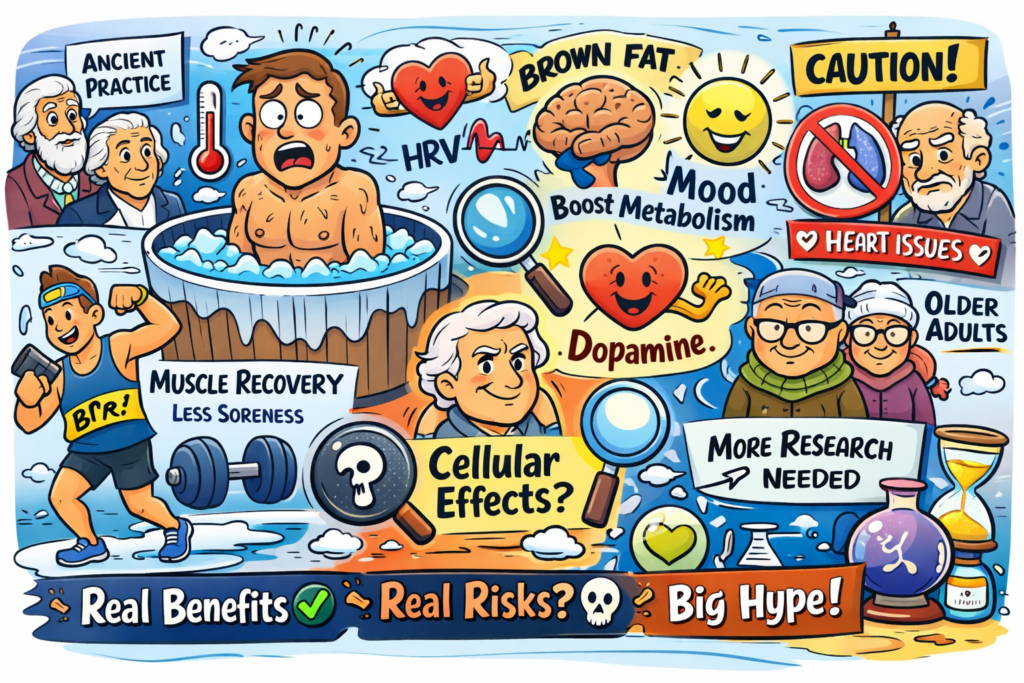


The One True Gospel of Wellness
By John Turley
On April 23, 2026
In Commentary, Medicine
Why Every Guru Thinks They’ve Found the Only Path to Health
There’s a peculiar affliction that seems to strike fitness influencers, biohackers, homeopathic healers, and wellness gurus with near-universal consistency — the unshakeable conviction that they, and only they, have cracked the code on human health. Whether it’s cold plunges at 4 a.m., microdosing mushrooms, coffee enemas, or whatever supplement stack is trending this week, every one of these prophets arrives at the same conclusion: their method is the path, the others are at best misguided, and mainstream medicine is a corrupt temple worth burning down.
Psychologists have a name for part of what’s happening here. It’s called the Dunning-Kruger effect — the tendency for people with limited knowledge in a domain to overestimate their own competence. But that’s only part of the story. Many of these figures are genuinely smart, sometimes even credentialed. What really drives the zealotry is something closer to what researchers call “belief perseverance” — the tendency to hold tightly to a conclusion even when contradicting evidence rolls in. Once someone has built an identity, a brand, and an income stream around a single idea, the psychological and financial cost of admitting nuance becomes enormous.
Take the biohacking community as a prime example. Some influencers — like the self-proclaimed “father of biohacking” — have built empires on the premise that optimizing the body is a matter of finding the right levers and pulling them correctly. They have championed everything from Bulletproof Coffee to infrared saunas to testosterone replacement, positioning each as a revelation that conventional medicine is too slow or too corrupted to acknowledge. The problem isn’t that all of these interventions lack merit — some have legitimate science behind them. The problem is the rhetorical framework: the idea that skeptics aren’t just wrong, they’re complicit. That’s not science; that’s a revival meeting.
Homeopathy sits at a different extreme but runs on the same engine. Developed in the late 18th century by Samuel Hahnemann, homeopathy is based on the idea that substances that cause symptoms in healthy people can cure those symptoms in the sick — and that extreme dilution actually strengthens a remedy’s potency. The scientific consensus is unambiguous: systematic reviews and meta-analyses have repeatedly found homeopathic remedies perform no better than placebo. And yet its advocates don’t merely disagree with this consensus — they dismiss the entire evidentiary framework, arguing that conventional research methods simply can’t measure what homeopathy does. It’s an airtight position: no evidence can ever count against it.
The fitness world runs its own version of this dogmatism on a perpetual loop. CrossFit devotees insist that anything other than functional high-intensity training is a waste of time. Carnivore diet advocates declare that vegetables are quietly poisoning you with antinutrients. Yoga instructors sometimes slide into the claim that breath control and mindfulness can substitute for actual medical care. Each subculture has its orthodoxy, its apostles, and its convenient explanations for why people who don’t follow the program are sick, lazy, or deceived. The irony is that many of these systems contain genuinely useful elements. Resistance training really does build muscle and bone density. Mindfulness really does reduce cortisol. Dietary quality really does matter enormously. But the insistence on one method to the exclusion of all others transforms useful practices into something closer to religious doctrine.
What’s lost in all the noise is the most important truth in medicine: human bodies are wildly heterogeneous. What works beautifully for one person may be ineffective or even harmful for another. This isn’t a flaw in the science — it is the science. Precision medicine, one of the most promising frontiers in modern healthcare, is built entirely on this recognition. The dream of a single universal protocol for human health isn’t just unrealized — it’s probably unrealizable. Yet that’s precisely what every wellness guru is selling.
There’s also a social dimension worth naming. The wellness industry is, in the most literal sense, an industry. It generated an estimated $5.6 trillion globally in 2022, according to the Global Wellness Institute, and that number continues to climb. When someone’s livelihood depends on their particular system being not just good but uniquely correct, objectivity becomes a luxury they can’t easily afford. Dismissing alternatives isn’t just tribalism — it’s good business.
None of this is to say that skepticism toward mainstream medicine is always misplaced. Conventional healthcare has real blind spots — in chronic disease management, in nutrition research, in the treatment of pain, and in its historical tendency to dismiss patient experience. The gurus often fill genuine gaps that the system has left open. But filling a gap is different from claiming you have the only map to the entire territory. The honest answer in health and fitness, as in most complex domains, is that we know a good deal, we don’t know quite enough, and anyone who tells you they’ve figured it all out probably hasn’t.
The next time someone tells you they’ve discovered the only way — whether it’s a supplement protocol, a spiritual practice, or a morning routine — it might be worth asking the simplest question in science: compared to what? If the answer is a dismissive wave at everything else, you probably have your answer.
Illustration generated by author using ChatGPT.
Sources
Global Wellness Institute — Global Wellness Economy Monitor: https://globalwellnessinstitute.org/industry-research/
Ernst E. — Homeopathy: The Undiluted Facts (Springer, 2016): https://link.springer.com/book/10.1007/978-3-319-43592-3
Dunning D. — The Dunning-Kruger Effect, Advances in Experimental Social Psychology: https://www.sciencedirect.com/science/article/pii/S0065260111440024
National Institutes of Health — Precision Medicine Initiative: https://www.nih.gov/research-training/allofus-research-program
Medical Disclaimer
The information provided in this article is intended for general educational and informational purposes only and does not constitute medical advice. It should not be used as a substitute for professional medical advice, diagnosis, or treatment.
Always seek the guidance of a qualified healthcare provider with any questions you may have regarding a medical condition or treatment. Never disregard professional medical advice or delay seeking it because of something you have read here.
If you are experiencing a medical emergency, call 911 or your local emergency number immediately.
The author of this article is a licensed physician, but the views expressed here are solely those of the author and do not represent the official position of any hospital, health system, or medical organization with which the author may be affiliated.