Walk through the supplement aisle of any pharmacy and you’ll find shelf after shelf of promises — stronger bones, sharper memory, less joint pain, better sleep. Americans spend roughly $60 billion a year on dietary supplements, and seniors are among the most enthusiastic buyers. But which of these products actually deliver, which are harmless but ineffective, and which could do real damage? The answers are more nuanced than the marketing suggests.
Older adults are often drawn to supplements because aging changes appetite, digestion, medication use, and nutrient absorption. But the general rule is simple: supplements work best when they fill a documented gap, and they are least useful when they are taken as a broad “insurance policy” by otherwise well-nourished people. Let’s take a closer look.
First, a ground rule that applies to everything in this article: dietary supplements are not FDA-approved drugs. The FDA treats them more like foods, meaning manufacturers don’t have to prove effectiveness before selling them. Quality control also varies widely — what’s on the label may not always match what’s in the bottle. As a result, the scientific evidence behind many supplements is limited or inconsistent. When shopping, look for products with a USP (United States Pharmacopeia) verified mark, which indicates independent testing for identity, purity, and potency.
The Genuinely Helpful Ones
Vitamin D and Calcium are probably the most well-supported supplements for older adults. Bone loss accelerates with age, and these two nutrients work as a team — calcium provides the raw material for bone, while vitamin D helps the body absorb it. The National Institute on Aging recommends 600 IU of vitamin D daily for adults aged 51–70, and 800 IU for those over 70. Most seniors don’t get enough from diet or sun exposure alone, making supplementation genuinely sensible for many people. This is especially true for prople with documented deficiency or osteoporosis risk. One important caveat: don’t go overboard. Too much vitamin D can cause calcium to build up in the blood, potentially harming the kidneys and blood vessels.
Vitamin B12 is another legitimate priority; Up to 15 percent of older adults may bedeficient.. Older adults are prone to B12 deficiency not because they eat less of it, but because the stomach produces less acid with age, and stomach acid is needed to release B12 from food. Those taking acid-blocking medications are at even higher risk. Deficiency can cause nerve damage and anemia. The good news is that the form of B12 in supplements is absorbed without needing stomach acid, making supplements effective where food sources may fall short.
Omega-3 fatty acids, found in fish oil, have earned a solid reputation for lowering triglycerides — a type of blood fat linked to heart disease. A large study of over 400,000 people found associations between fish oil use and improved cholesterol profiles. However, the picture is more complicated for other claimed benefits. Evidence for omega-3s preventing dementia is mixed, and some research suggests fish oil can actually raise LDL (“bad”) cholesterol in certain people, so monitoring is wise. For those who can’t eat fatty fish regularly, fish oil is a reasonable backup — just don’t expect miracles beyond the triglyceride benefit.
Melatonin has moderate scientific support for improving sleep, which is a chronic issue for many older adults. It’s particularly helpful for resetting disrupted sleep cycles. The key is using it at low doses — often 0.5 to 3 mg is sufficient, though most over-the-counter products contain far more. It’s generally well tolerated but should not replace evaluation of underlying sleep disorders.
Creatine and protein supplements may sound like something only gym rats need, but research increasingly supports their role in combating sarcopenia — the age-related loss of muscle mass that can lead to falls and loss of independence. A 2024 Stanford review found that creatine supplementation, combined with resistance training, can meaningfully preserve muscle in adults over 65. Branched-chain amino acids (BCAAs) can play a supporting role in certain situations, particularly when protein intake from food is inadequate. Vegans should pay particular attention to protein intake.
The Ambiguous Middle Ground
Glucosamine and chondroitin are among the most popular supplements for joint pain, and the scientific debate around them has been going on for decades. These are naturally occurring compounds in cartilage, and the theory is that supplementing them may slow joint deterioration in osteoarthritis. A 2024 systematic review of 146 studies found that over 90% of the studies reported positive outcomes — impressive on its face. But the landmark NIH-funded GAIT trial told a more sobering story: glucosamine and chondroitin, alone or together, were no more effective than a placebo for most people with knee osteoarthritis. The exception was a subgroup with moderate-to-severe pain, who did show moderate improvement. Safety is generally good, but those on blood thinners like warfarin should be careful, as glucosamine may affect clotting.
Turmeric and curcumin have generated enormous popular interest, and there’s at least a plausible scientific basis for the excitement. Curcumin, the active compound in turmeric, is a potent anti-inflammatory and antioxidant. Multiple clinical trials support some benefit for knee pain, and some research suggests potential benefits for cognitive health. However, curcumin is poorly absorbed on its own, which is why many products add black pepper (piperine) or use enhanced delivery formulations. The overall evidence, while promising, is still described as “mixed or low quality” by most reviewers. If you do try it, look for a formulation with enhanced bioavailability and give it at least 4–8 weeks and be aware that it may cause gastrointestinal symptoms.
Saw palmetto is widely used by older men for symptoms of benign prostatic hyperplasia (BPH) — the enlarged prostate that causes frequent urination. A 2024 updated Cochrane review found some evidence of limited benefit for urinary symptoms for some men, though the results are inconsistent and most mainstream urology guidelines do not formally recommend it. It’s generally well tolerated. Men using it should still get their prostate checked regularly and not assume saw palmetto rules out other conditions.
Magnesium has had a social media moment, with enthusiastic claims about better sleep, improved mood, and reduced muscle cramps. The actual science is more cautious — there’s limited evidence for magnesium supplements providing any of these benefits in people who aren’t already deficient. That said, deficiency is relatively common in older adults, and correction of a true deficiency can absolutely help. A blood test can tell you if you actually need it.
Multivitamins present a genuine paradox. They’re the most commonly taken supplement category, often recommended by physicians as a nutritional safety net. And for seniors with reduced appetite or limited dietary variety, that logic holds. But large, well-designed studies have found limited evidence that multivitamins improve longevity or prevent major diseases in otherwise healthy older adults. A newer 2024 analysis from the COSMOS trial suggests some modest benefit for cognitive function. Senior-specific multivitamins are preferred — they typically contain more vitamin D and B12 and less or no iron, which reflects the actual needs of older adults.
The Ones That Raise Red Flags
Iron supplements deserve special caution in older men and post-menopausal women. Unless there’s a documented deficiency confirmed by blood testing, taking iron supplements can be harmful. In men, iron overload is a genuine risk, and about twice as many men carry the gene for hereditary hemochromatosis (a condition where the body absorbs too much iron) as carry the gene for iron deficiency. Excess iron has been linked to liver damage and may raise cancer risk. Senior-specific multivitamins wisely contain little or no iron for exactly this reason.
High-dose Vitamin A is another potential problem. The liver’s ability to clear vitamin A decreases with age, and older adults absorb more of it. Doses above recommended daily values can accumulate to toxic levels, potentially harming the liver. This is specifically the retinol form of vitamin A. Beta-carotene from plant sources is much safer. Check your multivitamin label carefully.
High dose Vitamin B6 can cause nerve damage, balance problems, and sensory neuropathy when taken over long periods but is safe at recommended levels.
Many supplements claim to improve memory or prevent dementia. Unfortunately, the evidence is generally weak. Fish oil, ginkgo biloba, and other popular products have not demonstrated clear benefits for preventing cognitive decline in controlled studies. Some research suggests that long-term supplementation with B vitamins might slow certain aspects of cognitive decline in specific populations, but results remain inconsistent.
St. John’s Wort is widely used for mild depression, but it comes with a serious warning: it interacts with a long list of medications, including antidepressants, blood thinners, heart medications, and antiretroviral drugs. For seniors managing multiple conditions with multiple prescriptions, this herb is particularly risky. Ginkgo biloba carries similar drug interaction concerns, especially around bleeding risk when combined with blood thinners or aspirin.
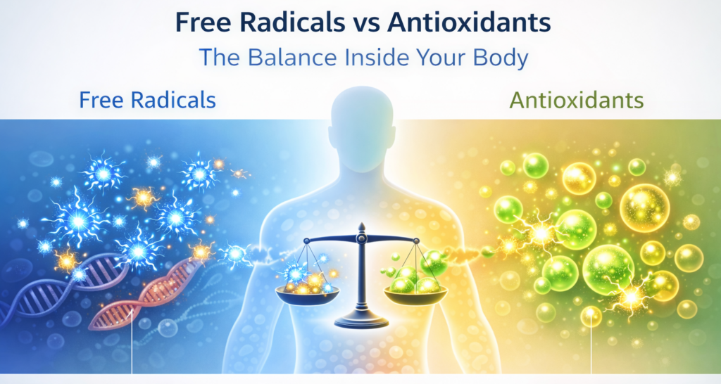
High-dose antioxidants — vitamins A, C, and E taken in large amounts — have largely failed to deliver on their promise of preventing heart disease and cancer. The US Preventive Services Task Force does not recommend these for prevention. In some cases, large antioxidant supplements may actually interfere with the body’s natural disease-fighting mechanisms.
The Bottom Line
Given the mixed evidence, a sensible approach to supplements includes several principles:
- Food first. A balanced diet usually provides most necessary nutrients.
- Test before supplementing. Blood tests can identify deficiencies such as B12 or Vitamin D.
- Avoid megadoses. Excessive intake of vitamins can cause toxicity.
- Check medication interactions. Many supplements interact with common drugs, including blood thinners.
- Treat supplements like medications. They should have a clear purpose and measurable benefit.
Supplements that address documented deficiencies or fill genuine dietary gaps — vitamin D, B12, calcium, omega-3s — offer the best evidence for benefit in seniors. Joint supplements like glucosamine and turmeric may help some people, though the evidence is mixed enough that a try-and-see approach (with a 2–3 month window to assess benefit) is reasonable. And several common supplements, particularly iron in unsupervised use, high-dose vitamin A, and certain herbals in combination with medications, carry risks that are easy to overlook because they’re sold without a prescription.
I always advised my patients to bring all their supplement bottles to at least one visit each year and to bring any medicines prescribed by specialists. Physicians can spot dangerous overlaps, flag interactions with your prescriptions, and tell you if what you’re taking makes sense for you. Many seniors never hear a list of side effects for supplements the way they do for prescription drugs — and they often assume that means there aren’t any. That assumption, unfortunately, can be costly.
Illustration generated by author using ChatGPT.
Sources
National Institute on Aging. Dietary Supplements for Older Adults.
National Institute on Aging. Vitamins and Minerals for Older Adults.
Linus Pauling Institute, Oregon State University. Older Adults — Micronutrient Information Center.
National Center for Health Research. Glucosamine Supplements: Do They Work and Are They Safe?
BodySpec. Supplements for Joint Health: 2025 Evidence-Based Guide.
Cleveland Clinic. Dietary Supplements Compound Health Issues for Older Adults.
FDA. Mixing Medications and Dietary Supplements Can Endanger Your Health.
NIH Office of Dietary Supplements. Iron — Health Professional Fact Sheet.
Foods (MDPI). Food Supplements and Their Use in Elderly Subjects — Challenges and Risks. 2024.
Memorial Healthcare System. Herbal Supplements and Prescription Drugs: Know the Risks. 2024.
WebMD. Saw Palmetto: Overview, Uses, Side Effects, Precautions.
________________________________________________
Medical Disclaimer
The information provided in this article is intended for general educational and informational purposes only and does not constitute medical advice. It should not be used as a substitute for professional medical advice, diagnosis, or treatment.
Always seek the guidance of a qualified healthcare provider with any questions you may have regarding a medical condition or treatment. Never disregard professional medical advice or delay seeking it because of something you have read here.
If you are experiencing a medical emergency, call 911 or your local emergency number immediately.
The author of this article is a licensed physician, but the views expressed here are solely those of the author and do not represent the official position of any hospital, health system, or medical organization with which the author may be affiliated.