Grumpy opinions about everything.

Tag: Medical Technology

Seeing Inside: A Guide to Modern Diagnostic Radiology

Not so long ago, if a doctor needed to know what was happening inside your body, the options were limited — a basic X-ray, an exploratory surgery, or educated guesswork. Today, a radiologist can map your brain’s blood vessels, detect a cancer smaller than a pea, or watch your heart metabolizing sugar in near real time — all without making a single incision.

The first medical X-ray appeared in 1895. For decades, imaging largely meant detecting fractures or large abnormalities. Today, radiology guides emergency care, cancer treatment, stroke therapy, cardiac management, and preventive medicine. Modern diagnostic imaging has transformed medicine in ways that would have seemed like science fiction to physicians just a few generations ago.

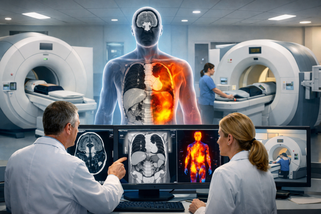
Modern imaging falls into three broad categories: structural imaging — what tissues look like, vascular imaging — how blood flows, and functional imaging — how cells behave metabolically. Here’s a plain-language guide to the big three: MRI/MRA, CT/CTA, and PET scans — what they are, how they work, and why they matter.

MRI and MRA: Magnets and Radio Waves

The MRI — magnetic resonance imaging — is one of the most versatile tools in modern medicine, and it works without a single ray of radiation. An MRI passes an electric current through coiled wires to create a temporary magnetic field in your body. A transmitter and receiver then send and receive radio waves, and a computer uses those signals to construct detailed digital images of whatever area is being scanned. Think of it as a very sophisticated tuning fork: it causes hydrogen atoms in your body’s water molecules to briefly align, then releases them — and the energy they emit on the way back creates the image. Because different tissues relax at different rates, MRI can distinguish gray matter from white matter in the brain, normal from inflamed or cancerous tissue, and ligament from muscle with impressive contrast.

The result is exceptional detail, especially for soft tissue. MRI scans take much clearer pictures of your brain, spinal cord, nerves, muscles, ligaments, and tendons than regular X-rays and CT scans. That’s why your orthopedic surgeon orders one when your knee goes sideways, and why neurologists reach for it when they suspect a stroke or multiple sclerosis.

MRA — magnetic resonance angiography — is MRI’s cousin, using the same magnetic technology but focused specifically on blood vessels. It lets physicians map arteries and veins in remarkable detail, identifying narrowing (stenosis), bulges (aneurysms), or blockages (occlusions) without the need for invasive catheterization. If your doctor suspects a blockage in the blood vessels feeding your brain or kidneys, an MRA can reveal it clearly. A contrast dye is sometimes injected to make vessels stand out even more sharply.

The main trade-offs with MRI are time and noise — scans generally take between 30 to 50 minutes, and the machine produces the kind of clanging racket that makes earplugs standard issue. People with certain metal implants or severe claustrophobia can’t always use it, which is where CT steps in.

CT and CTA: X-Rays, Upgraded

The CT scan — computed tomography — takes the familiar chest X-ray and turns it into something far more powerful. A CT scan takes multiple X-ray images from different angles rotating around the body, separates them by depth then processes them by computer to create cross-sectional views — essentially a detailed 3D picture rather than a flat 2D image. Think of slicing a loaf of bread: instead of seeing only the crust, you can examine every slice.

A CT shows more detail than a standard X-ray and is used to diagnose cancer, heart disease, injuries from trauma, and musculoskeletal disorders — it’s one of the most common imaging tests used today.  Emergency departments rely on CT heavily because it’s fast, often completed in 10 to 15 minutes, and can quickly identify life-threatening conditions like internal bleeding or pulmonary embolism.

CTA — CT angiography — adds an injected contrast dye to the mix, allowing physicians to see blood vessels with high clarity. Contrasting agents help show various structures of the body more clearly, making CTA the go-to test for evaluating coronary arteries, aortic aneurysms, and vascular disease throughout the body. Unlike MRA, it’s faster and more widely available, though it does involve a dose of radiation — something physicians weigh carefully against the diagnostic benefit.

PET Scan: When Function Matters More Than Form

If MRI and CT show you the structure of the body, the PET scan — positron emission tomography — shows you what’s happening inside it. This is a fundamentally different question, and the technology reflects that.

A PET scan is used to see metabolism and chemical activity within your body. It can detect abnormal changes before structural changes occur — meaning it can detect cancer before a tumor is large enough to be seen on a CT or MRI. That’s a remarkable capability. The scan works by injecting a small amount of a radioactive tracer — most commonly a form of glucose — into your bloodstream. Diseased cells, particularly cancer cells, absorb more of the radiotracer than healthy ones do.  Active cells consume glucose. Cancer cells, inflamed tissue, and active brain regions often use more glucose than surrounding tissue. These are called “hot spots,” and the PET scanner detects this radiation to produce images of affected tissue.

Beyond cancer, PET scans are invaluable in cardiology — showing whether heart muscle is still alive after a heart attack — and in neurology, helping diagnose Alzheimer’s disease, epilepsy, and brain tumors by revealing abnormal patterns of brain activity.

The trade-off is resolution. The image resolution of nuclear medicine images may not be as high as that of CT or MRI, which is why PET is rarely used alone today. Combined PET/CT scanners perform almost all PET scans today , marrying metabolic information with anatomical precision. Hybrid PET/MRI scanners are also emerging though not yet in widespread use. They are particularly valuable for soft-tissue cancers of the brain, liver, and pelvis.

How They Compare — and Why It Matters

The simplest way to think about these three technologies is this: CT shows shape and structure quickly, with emphasis on bone and dense tissue; MRI shows soft tissue in extraordinary detail without radiation; and PET shows function and cellular activity that neither of the others can see directly.

A patient with suspected cancer might undergo a CT scan to identify the location of a mass, followed by a PET to determine its metabolic activity, and then an MRI to map its relationship to critical soft tissues. Used together, these tools give physicians a picture of disease that is more complete than any single test could provide.

The value to modern medicine is difficult to overstate. These technologies allow doctors to diagnose conditions earlier, stage cancers more accurately, guide surgical planning, and monitor how well treatments are working — all without exploratory surgery. Survival rates for many cancers have improved substantially in part because imaging lets us find disease when it’s still manageable.

The era when medicine was largely guesswork about what lay beneath the skin is over. Today, radiologists are, in a very real sense, reading the body like an open book.

Illustration generated by author using ChatGPT.

Sources:

UNC Health Appalachian — MRI, CT, and PET Scan Comparison: https://www.unchealthappalachian.org/blog/2024/comparing-mri-ct-and-pet-scans-how-they-work-and-when-theyre-use/

WashU Mallinckrodt Institute of Radiology — Differences Between CT, MRI, and PET: https://www.mir.wustl.edu/do-you-know-the-differences-between-a-ct-mri-and-pet-scan/

Cleveland Clinic — PET Scan Overview: https://my.clevelandclinic.org/health/diagnostics/10123-pet-scan

RadiologyInfo.org — PET/CT: https://www.radiologyinfo.org/en/info/pet

Open MedScience — CT, MRI, and PET Differences: https://openmedscience.com/ct-mri-and-pet-scanners-unravelling-the-differences-in-modern-medical-imaging/

Healthline — MRI vs. PET Scan: https://www.healthline.com/health/mri-vs-pet-scan

Revere Health — MRI, CT, and PET Explained: https://reverehealth.com/live-better/mri-ct-pet/

Fecal Microbiota Transplantation: When Waste Becomes Therapy

Today I’m going to talk about something that may sound unbelievable and maybe even a little gross—fecal transplant. Yes, it’s exactly what it sounds like. Getting a transplant of someone else’s poop.

The human gut contains trillions of microorganisms—bacteria, viruses, fungi—living in a complex ecosystem that influences everything from digestion to immune function. This is called the microbiome.  When this ecosystem gets disrupted, the consequences can range from uncomfortable to life-threatening. Enter one of medicine’s most counterintuitive treatments: fecal microbiota transplantation, or FMT, where stool from a healthy donor is transferred to a patient to restore a healthy community of gut microbes.

What Is FMT

The basic idea is simple: if someone’s microbiome has been badly disrupted (most commonly by repeated antibiotic exposure), replacing it with a balanced microbial ecosystem can help the gut recover.  At its core, FMT is taking fecal matter from a healthy donor and introducing it into a patient’s gastrointestinal tract. But it’s not the solid waste itself that matters; it’s the billions of beneficial bacteria and other microorganisms living in that material. Think of it as a probiotic treatment on steroids, delivering an entire functioning ecosystem rather than just a few select bacterial strains.

The gut microbiome plays crucial roles in digestion, vitamin production, immune system regulation, and even protection against harmful pathogens. When antibiotics, illness, or other factors devastate this ecosystem, dangerous bacteria like Clostridioides difficile (C. diff) can take over, causing severe diarrhea, inflammation, and potentially fatal infections.

The Clinical Track Record

While it may sound like “weird science”, FMT has been around for centuries. It was used in ancient Chinese medicine in a formulation called “yellow soup“ to treat food poisoning and intractable diarrhea. It was used as early as the 16th century in Europe to treat sick farm animals, particularly sheep and cattle.

FMT’s most dramatic success story involves C. diff infections, particularly the recurrent cases that don’t respond to antibiotics. Multiple randomized controlled trials have shown FMT to be remarkably effective—with cure rates often exceeding 80-90% for recurrent C. diff infections, compared to roughly 25-30% for continued antibiotic therapy. A landmark 2013 study reported in the New England Journal of Medicine was stopped early because FMT was so dramatically superior to standard treatment that continuing to withhold it from the control group seemed unethical.

Beyond C. diff, researchers are investigating FMT for inflammatory bowel diseases like ulcerative colitis and Crohn’s disease, with mixed but occasionally promising results. Some studies have shown potential for ulcerative colitis, with remission rates around 24-27%. The research into Crohn’s disease, irritable bowel syndrome, metabolic disorders, and even neurological conditions is ongoing but less conclusive. The FDA currently considers FMT an investigational treatment for most conditions except recurrent C. diff, where it’s become a recognized therapeutic option.

How It Works

The actual process of FMT can use several routes. The most common approaches involve colonoscopy, where the donated material is delivered directly to the colon, or through nasogastric or nasoduodenal tubes that thread through the nose down to the small intestine. More recently, oral capsules containing frozen, encapsulated donor stool have become available, offering a less invasive alternative that patients often prefer.

Before the transplant, the donated stool is carefully processed. It’s typically mixed with a saline solution and filtered to remove large particles while preserving the microbial communities. The resulting liquid suspension is what gets delivered to the patient. For frozen preparations, this material is mixed with a cryoprotectant, frozen at extremely cold temperatures, and can be stored for months before use.

The preparation isn’t just about the donor material—patients often undergo their own preparation. Many protocols include antibiotics to reduce the overgrowth of harmful bacteria before the transplant, followed by bowel cleansing similar to what you’d do before a colonoscopy. The idea is to create a relatively clean slate where the new microbial ecosystem can establish itself.

Sources of Donor Material

This brings us to one of the most critical aspects: donor selection and screening. Not just anyone can donate stool for medical use. The screening process is extensive and rigorous, rivaling or exceeding the scrutiny applied to blood donation.

Donors undergo detailed health questionnaires covering everything from recent travel and antibiotic use to gastrointestinal symptoms and risk factors for infectious diseases. They provide blood and stool samples that are tested for a long list of potential pathogens: C. diff, Helicobacter pylori, parasites, hepatitis A, B, and C, HIV, syphilis, and various other bacteria and viruses. The FDA issued guidance requiring additional testing for multi-drug resistant organisms after several patients contracted serious infections from FMT.

Donors generally fall into two categories: directed donors and universal donors. Directed donors are typically family members or friends who undergo screening and provide stool specifically for one patient. Universal donors go through the same rigorous screening but provide samples that can be used for multiple patients. These universal donors often work with stool banks—specialized facilities that collect, process, screen, and distribute donor material to healthcare providers.

The largest stool bank in the United States, OpenBiome, was founded in 2012 and has processed material from thousands of donors for tens of thousands of treatments. They report that only about 2-3% of volunteer donors successfully make it through the screening process, highlighting just how selective the criteria are. These banks have made FMT more widely available, eliminating the need for individual healthcare facilities to find and screen their own donors.

The Balance of Promise and Caution

While FMT represents a genuine breakthrough for recurrent C. diff infections, the medical community remains appropriately cautious about expanding its use. The FDA regulates FMT and has expressed concerns about potential risks, particularly after cases where patients developed serious infections from inadequately screened donors. There questions about the long-term effects of introducing another person’s microbiome, and there are theoretical concerns about transmitting conditions or predispositions we don’t fully understand.

The research into FMT for conditions beyond C. diff continues, but many trials have shown modest or inconsistent results. The microbiome’s role in health and disease is incredibly complex, and what works dramatically for one condition may not translate to others. Still, the fundamental insight—that our gut microbiome profoundly influences our health and that we can therapeutically manipulate it—has opened potential new avenues in medicine.

Sources

                1. van Nood, E., et al. (2013). “Duodenal Infusion of Donor Feces for Recurrent Clostridium difficile.” New England Journal of Medicine, 368(5), 407-415. https://www.nejm.org/doi/full/10.1056/NEJMoa1205037

                2. U.S. Food and Drug Administration. “Fecal Microbiota for Transplantation: Safety Information.” https://www.fda.gov/vaccines-blood-biologics/safety-availability-biologics/fecal-microbiota-transplantation-safety-information

                3. Cammarota, G., et al. (2017). “European consensus conference on faecal microbiota transplantation in clinical practice.” Gut, 66(4), 569-580. https://gut.bmj.com/content/66/4/569

                4. Moayyedi, P., et al. (2015). “Fecal Microbiota Transplantation Induces Remission in Patients With Active Ulcerative Colitis in a Randomized Controlled Trial.” Gastroenterology, 149(1), 102-109. https://www.gastrojournal.org/article/S0016-5085(15)00381-5/fulltext

                5. Kelly, C.R., et al. (2016). “Update on Fecal Microbiota Transplantation 2015: Indications, Methodologies, Mechanisms, and Outlook.” Gastroenterology, 150(1), 276-290. https://www.gastrojournal.org/article/S0016-5085(15)01626-7/fulltext

                6. OpenBiome. “Our Process: Screening.” https://www.openbiome.org/safety

                7. Quraishi, M.N., et al. (2017). “Systematic review with meta-analysis: the efficacy of faecal microbiota transplantation for the treatment of recurrent and refractory Clostridium difficile infection.” Alimentary Pharmacology & Therapeutics, 46(5), 479-493. https://onlinelibrary.wiley.com/doi/full/10.1111/apt.14201​​​​​​​​​​​​​​​​

Illustration generated by author using Midjourney

Powered by WordPress & Theme by Anders Norén